

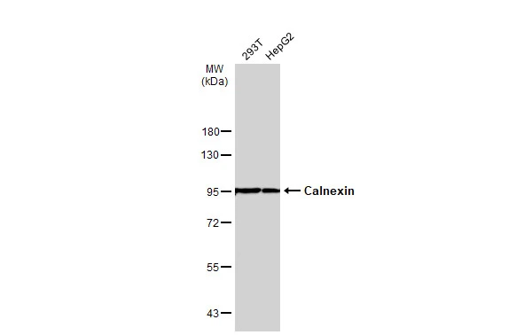
Various whole cell extracts (30 μg) were separated by 7.5% SDS-PAGE, and the membrane was blotted with Calnexin antibody [N3C2], Internal (GTX101676) diluted at 1:5000. The HRP-conjugated anti-rabbit IgG antibody (GTX213110-01) was used to detect the primary antibody.
![Calnexin antibody [N3C2], Internal detects Calnexin protein at cytoplasm by immunohistochemical analysis. Sample: Paraffin-embedded rat testis. Calnexin stained by Calnexin antibody [N3C2], Internal (GTX101676) diluted at 1:500. Antigen Retrieval: Citrate buffer, pH 6.0, 15 min](https://www.genetex.com/upload/website/prouct_img/normal/GTX101676/GTX101676_43644_20190830_IHC-P_R_w_23060100_511.webp "Calnexin antibody [N3C2], Internal detects Calnexin protein at cytoplasm by immunohistochemical analysis. Sample: Paraffin-embedded rat testis. Calnexin stained by Calnexin antibody [N3C2], Internal (GTX101676) diluted at 1:500. Antigen Retrieval: Citrate buffer, pH 6.0, 15 min")
![Non-transfected (–) and transfected (+) 293T whole cell extracts (15 μg) were separated by 7.5% SDS-PAGE, and the membrane was blotted with Calnexin antibody [N3C2], Internal (GTX101676) diluted at 1:10000. The HRP-conjugated anti-rabbit IgG antibody (GTX213110-01) was used to detect the primary antibody.](https://www.genetex.com/upload/website/prouct_img/normal/GTX101676/GTX101676_39897_20160707_WB_shRNA_watermark_w_23060100_140.webp "Non-transfected (–) and transfected (+) 293T whole cell extracts (15 μg) were separated by 7.5% SDS-PAGE, and the membrane was blotted with Calnexin antibody [N3C2], Internal (GTX101676) diluted at 1:10000. The HRP-conjugated anti-rabbit IgG antibody (GTX213110-01) was used to detect the primary antibody.")
 A:NIH-3T3 7.5% SDS PAGE GTX101676 diluted at 1:1000 The HRP-conjugated anti-rabbit IgG antibody (GTX213110-01) was used to detect the primary antibody.")
![Calnexin antibody [N3C2], Internal detects Calnexin protein at cytoplasm by immunohistochemical analysis. Sample: Paraffin-embedded mouse liver. Calnexin stained by Calnexin antibody [N3C2], Internal (GTX101676) diluted at 1:500. Antigen Retrieval: Citrate buffer, pH 6.0, 15 min](https://www.genetex.com/upload/website/prouct_img/normal/GTX101676/GTX101676_43645_20190830_IHC-P_M_w_23060100_856.webp "Calnexin antibody [N3C2], Internal detects Calnexin protein at cytoplasm by immunohistochemical analysis. Sample: Paraffin-embedded mouse liver. Calnexin stained by Calnexin antibody [N3C2], Internal (GTX101676) diluted at 1:500. Antigen Retrieval: Citrate buffer, pH 6.0, 15 min")
![Calnexin antibody [N3C2], Internal detects Calnexin protein at cytoplasm by immunohistochemical analysis. Sample: Paraffin-embedded rat kidney. Calnexin stained by Calnexin antibody [N3C2], Internal (GTX101676) diluted at 1:500. Antigen Retrieval: Citrate buffer, pH 6.0, 15 min](https://www.genetex.com/upload/website/prouct_img/normal/GTX101676/GTX101676_43645_20190830_IHC-P_R_w_23060100_292.webp "Calnexin antibody [N3C2], Internal detects Calnexin protein at cytoplasm by immunohistochemical analysis. Sample: Paraffin-embedded rat kidney. Calnexin stained by Calnexin antibody [N3C2], Internal (GTX101676) diluted at 1:500. Antigen Retrieval: Citrate buffer, pH 6.0, 15 min")
![Calnexin antibody [N3C2], Internal detects Calnexin protein at cytoplasm by immunohistochemical analysis. Sample: Paraffin-embedded mouse duodenum. Calnexin stained by Calnexin antibody [N3C2], Internal (GTX101676) diluted at 1:500. Antigen Retrieval: Citrate buffer, pH 6.0, 15 min](https://www.genetex.com/upload/website/prouct_img/normal/GTX101676/GTX101676_43642_20190830_IHC-P_M_w_23060100_931.webp "Calnexin antibody [N3C2], Internal detects Calnexin protein at cytoplasm by immunohistochemical analysis. Sample: Paraffin-embedded mouse duodenum. Calnexin stained by Calnexin antibody [N3C2], Internal (GTX101676) diluted at 1:500. Antigen Retrieval: Citrate buffer, pH 6.0, 15 min")
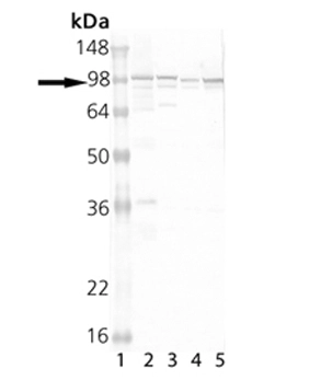
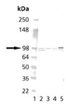
![Various tissue extracts (50 μg) were separated by 7.5% SDS-PAGE, and the membrane was blotted with Calnexin antibody [N3C2], Internal (GTX101676) diluted at 1:1000. The HRP-conjugated anti-rabbit IgG antibody (GTX213110-01) was used to detect the primary antibody.](https://www.genetex.com/upload/website/prouct_img/normal/GTX101676/GTX101676_43642_20220325_WB_M_R_w_23060100_521.webp "Various tissue extracts (50 μg) were separated by 7.5% SDS-PAGE, and the membrane was blotted with Calnexin antibody [N3C2], Internal (GTX101676) diluted at 1:1000. The HRP-conjugated anti-rabbit IgG antibody (GTX213110-01) was used to detect the primary antibody.")
![Calnexin antibody [N3C2], Internal detects Calnexin protein at cytoplasm by immunohistochemical analysis. Sample: Paraffin-embedded mouse testis. Calnexin stained by Calnexin antibody [N3C2], Internal (GTX101676) diluted at 1:500. Antigen Retrieval: Citrate buffer, pH 6.0, 15 min](https://www.genetex.com/upload/website/prouct_img/normal/GTX101676/GTX101676_43644_20190830_IHC-P_M_w_23060100_917.webp "Calnexin antibody [N3C2], Internal detects Calnexin protein at cytoplasm by immunohistochemical analysis. Sample: Paraffin-embedded mouse testis. Calnexin stained by Calnexin antibody [N3C2], Internal (GTX101676) diluted at 1:500. Antigen Retrieval: Citrate buffer, pH 6.0, 15 min")
![Calnexin antibody [N3C2], Internal detects Calnexin protein at cytoplasm by immunohistochemical analysis. Sample: Paraffin-embedded rat testis. Calnexin stained by Calnexin antibody [N3C2], Internal (GTX101676) diluted at 1:500. Antigen Retrieval: Citrate buffer, pH 6.0, 15 min](https://www.genetex.com/upload/website/prouct_img/normal/GTX101676/GTX101676_43642_20190830_IHC-P_R_w_23060100_255.webp "Calnexin antibody [N3C2], Internal detects Calnexin protein at cytoplasm by immunohistochemical analysis. Sample: Paraffin-embedded rat testis. Calnexin stained by Calnexin antibody [N3C2], Internal (GTX101676) diluted at 1:500. Antigen Retrieval: Citrate buffer, pH 6.0, 15 min")
Various whole cell extracts (30 μg) were separated by 7.5% SDS-PAGE, and the membrane was blotted with Calnexin antibody [N3C2], Internal (GTX101676) diluted at 1:5000. The HRP-conjugated anti-rabbit IgG antibody (GTX213110-01) was used to detect the primary antibody.
Calnexin antibody [N3C2], Internal
GTX101676
ApplicationsImmunoFluorescence, Western Blot, ImmunoCytoChemistry, ImmunoHistoChemistry, ImmunoHistoChemistry Paraffin
Product group Antibodies
ReactivityHuman, Mouse, Rat
TargetCANX
Overview
- SupplierGeneTex
- Product NameCalnexin antibody [N3C2], Internal
- Delivery Days Customer9
- Application Supplier NoteWB: 1:500-1:10000. ICC/IF: 1:100-1:1000. IHC-P: 1:100-1:1000. *Optimal dilutions/concentrations should be determined by the researcher.Not tested in other applications.
- ApplicationsImmunoFluorescence, Western Blot, ImmunoCytoChemistry, ImmunoHistoChemistry, ImmunoHistoChemistry Paraffin
- CertificationResearch Use Only
- ClonalityPolyclonal
- Concentration0.16 mg/ml
- ConjugateUnconjugated
- Gene ID821
- Target nameCANX
- Target descriptioncalnexin
- Target synonymsCNX, IP90, P90, calnexin, epididymis secretory sperm binding protein, major histocompatibility complex class I antigen-binding protein p88
- HostRabbit
- IsotypeIgG
- Protein IDP27824
- Protein NameCalnexin
- Scientific DescriptionThis gene encodes a member of the calnexin family of molecular chaperones. The encoded protein is a calcium-binding, endoplasmic reticulum (ER)-associated protein that interacts transiently with newly synthesized N-linked glycoproteins, facilitating protein folding and assembly. It may also play a central role in the quality control of protein folding by retaining incorrectly folded protein subunits within the ER for degradation. Alternatively spliced transcript variants encoding the same protein have been described. [provided by RefSeq]
- ReactivityHuman, Mouse, Rat
- Storage Instruction-20°C or -80°C,2°C to 8°C
- UNSPSC41116161
Datasheet
Related products
Product group Antibodies
Anti-Calnexin AntibodyA94879
ApplicationsImmunoFluorescence, Western Blot, ELISA, ImmunoHistoChemistry
ReactivityHuman, Mouse, Rat
- SizePrice

Product group Antibodies
Anti-Calnexin/CANX Antibody Picoband(r)A03372-2-CARRIER-FREE
ApplicationsFlow Cytometry, ImmunoFluorescence, Western Blot, ELISA, ImmunoCytoChemistry, ImmunoHistoChemistry
ReactivityHuman, Mouse
TargetCANX
- SizePrice
Product group Antibodies
Anti-CANX Antibody144-63846
ApplicationsImmunoFluorescence, Western Blot, ImmunoHistoChemistry
ReactivityHuman, Mouse, Rat
TargetCANX
- SizePrice
Product group Antibodies
CANX AntibodyCSB-PA001175
ApplicationsImmunoFluorescence, Western Blot, ELISA, ImmunoHistoChemistry
ReactivityHuman, Mouse, Rat
TargetCANX
- SizePrice
Product group Antibodies
References

Goat anti-CalnexinEB09525
ApplicationsImmunoFluorescence, Western Blot, ELISA, ImmunoHistoChemistry
ReactivityBovine, Canine, Human, Mouse, Porcine, Rat
TargetCANX
- SizePrice
Product group Antibodies
CANX Polyclonal AntibodyCAC14640
ApplicationsImmunoFluorescence, Western Blot, ELISA, ImmunoHistoChemistry
TargetCANX
- SizePrice
Product group Antibodies
References
Calnexin Polyclonal AntibodyBS-1693R
ApplicationsFlow Cytometry, ImmunoFluorescence, Western Blot, ELISA, ImmunoCytoChemistry, ImmunoHistoChemistry, ImmunoHistoChemistry Frozen, ImmunoHistoChemistry Paraffin
ReactivityBovine, Chicken, Equine, Human, Mouse, Rabbit, Rat
TargetCANX
- SizePrice
Product group Antibodies

CANX / Calnexin AntibodyLS-C401473
ApplicationsWestern Blot, ELISA, ImmunoHistoChemistry
ReactivityHuman, Mouse, Rat
TargetCANX
- SizePrice
Product group Antibodies
References
Calnexin antibodyGTX13504
ApplicationsFlow Cytometry, ImmunoFluorescence, ImmunoPrecipitation, Western Blot, ImmunoCytoChemistry, ImmunoHistoChemistry
ReactivityAvian, Bovine, Guinea Pig, Hamster, Human, Monkey, Mouse, Porcine, Rabbit, Rat, Sheep, Xenopus
TargetCANX
- SizePrice
Product group Antibodies
References
Calnexin antibodyGTX13505
ApplicationsFlow Cytometry, ImmunoFluorescence, ImmunoPrecipitation, Western Blot, ImmunoCytoChemistry, ImmunoHistoChemistry
ReactivityAvian, Bovine, Guinea Pig, Hamster, Human, Monkey, Mouse, Porcine, Rabbit, Rat, Sheep, Xenopus
TargetCANX
- SizePrice