

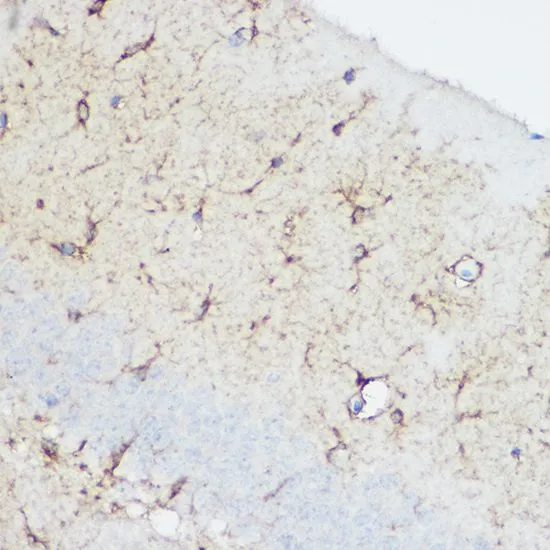
IHC-P analysis of mouse heart tissue using GTX32486 Calpain 1 antibody. Dilution : 1:100

IHC-P analysis of mouse heart tissue using GTX32486 Calpain 1 antibody. Dilution : 1:100
Calpain 1 antibody
GTX32486
ApplicationsWestern Blot, ImmunoHistoChemistry, ImmunoHistoChemistry Paraffin
Product group Antibodies
ReactivityHuman, Mouse
TargetCAPN1
Overview
- SupplierGeneTex
- Product NameCalpain 1 antibody
- Delivery Days Customer9
- Application Supplier NoteWB: 1:500 - 1:2000. IHC-P: 1:50 - 1:200. *Optimal dilutions/concentrations should be determined by the researcher.Not tested in other applications.
- ApplicationsWestern Blot, ImmunoHistoChemistry, ImmunoHistoChemistry Paraffin
- CertificationResearch Use Only
- ClonalityPolyclonal
- ConjugateUnconjugated
- Gene ID823
- Target nameCAPN1
- Target descriptioncalpain 1
- Target synonymsCANP, CANP1, CANPL1, SPG76, muCANP, muCL, calpain-1 catalytic subunit, CANP 1, calcium-activated neutral proteinase 1, calpain 1, (mu/I) large subunit, calpain mu-type, calpain, large polypeptide L1, calpain-1 large subunit, cell proliferation-inducing gene 30 protein, cell proliferation-inducing protein 30, micromolar-calpain
- HostRabbit
- IsotypeIgG
- Protein IDP07384
- Protein NameCalpain-1 catalytic subunit
- Scientific DescriptionThe calpains, calcium-activated neutral proteases, are nonlysosomal, intracellular cysteine proteases. The mammalian calpains include ubiquitous, stomach-specific, and muscle-specific proteins. The ubiquitous enzymes consist of heterodimers with distinct large, catalytic subunits associated with a common small, regulatory subunit. This gene encodes the large subunit of the ubiquitous enzyme, calpain 1. Several transcript variants encoding two different isoforms have been found for this gene. [provided by RefSeq, Nov 2010]
- ReactivityHuman, Mouse
- Storage Instruction-20°C or -80°C,2°C to 8°C
- UNSPSC41116161
Datasheet
Related products
Product group Antibodies
Anti-Calpain 1 AntibodyA83619
ApplicationsWestern Blot, ELISA, ImmunoHistoChemistry
ReactivityHuman
- SizePrice
Product group Antibodies
Anti-CAPN1 Antibody144-62123
ApplicationsWestern Blot, ImmunoHistoChemistry
ReactivityHuman, Mouse
TargetCAPN1
- SizePrice
Product group Antibodies
CAPN1 Recombinant Antibody, Biotin ConjugatedBSM-61500R-BIOTIN
ApplicationsWestern Blot, ImmunoHistoChemistry, ImmunoHistoChemistry Frozen, ImmunoHistoChemistry Paraffin
ReactivityHuman, Mouse, Rat
TargetCAPN1
- SizePrice
Product group Antibodies
CAPN1 AntibodyCSB-PA004490EA01HU
ApplicationsWestern Blot, ELISA, ImmunoHistoChemistry
ReactivityHuman
TargetCAPN1
- SizePrice
Product group Antibodies

Goat anti-CAPN1EB11988
ApplicationsWestern Blot, ELISA, ImmunoHistoChemistry
ReactivityHuman
TargetCAPN1
- SizePrice
Product group Antibodies
Capn1 Polyclonal AntibodyCAC07490
ApplicationsImmunoFluorescence, Western Blot, ELISA, ImmunoHistoChemistry
TargetCAPN1
- SizePrice
Product group Antibodies
Calpain 1 antibody [15C10]GTX16680
ApplicationsImmunoPrecipitation, Western Blot, ELISA
ReactivityBovine, Human, Mouse, Rat
TargetCAPN1
- SizePrice
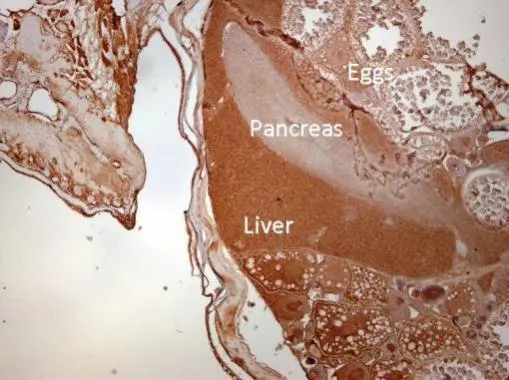
![Calpain 1 antibody [N3C2], Internal detects Calpain 1 protein at cytoplasm on mouse lung by immunohistochemical analysis. Sample: Paraffin-embedded mouse lung. Calpain 1 antibody [N3C2], Internal (GTX102340) diluted at 1:500.
Antigen Retrieval: Trilogy? (EDTA based, pH 8.0) buffer, 15min](https://www.genetex.com/upload/website/prouct_img/normal/GTX102340/GTX102340_40302_20150313_IHC_M_w_23060100_827.webp)
Product group Antibodies
Calpain 1 antibody [N3C2], InternalGTX102340
ApplicationsImmunoFluorescence, ImmunoPrecipitation, Western Blot, ImmunoCytoChemistry, ImmunoHistoChemistry, ImmunoHistoChemistry Paraffin
ReactivityHuman, Mammals, Mouse
TargetCAPN1
- SizePrice
Product group Antibodies
Calpain 1 antibodyGTX102350
ApplicationsFlow Cytometry, ImmunoFluorescence, Western Blot, ImmunoCytoChemistry, ImmunoHistoChemistry, ImmunoHistoChemistry Paraffin
ReactivityHuman, Zebra Fish
TargetCAPN1
- SizePrice

Product group Antibodies
Anti-CAPN1 AntibodyHPA005992
ApplicationsWestern Blot, ImmunoCytoChemistry, ImmunoHistoChemistry
ReactivityHuman
TargetCAPN1
- SizePrice