

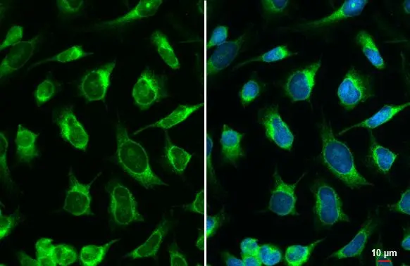
Calreticulin antibody detects Calreticulin protein at endoplasmic reticulum by immunofluorescent analysis. Sample: HeLa cells were fixed in ice-cold MeOH for 5 min. Green: Calreticulin stained by Calreticulin antibody (GTX111627) diluted at 1:500.
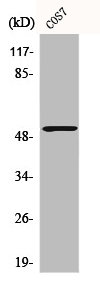
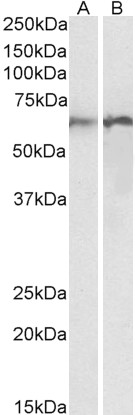
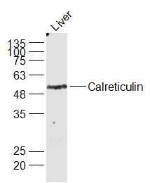
 were separated by 10% SDS-PAGE, and the membrane was blotted with Calreticulin antibody (GTX111627) diluted at 1:1000. The HRP-conjugated anti-rabbit IgG antibody (GTX213110-01) was used to detect the primary antibody.")
 antibody at 1:500 dilution.
Antigen Retrieval: Trilogy? (EDTA based, pH 8.0) buffer, 15min")
 antibody at 1:500 dilution.
Antigen Retrieval: Trilogy? (EDTA based, pH 8.0) buffer, 15min")
 were separated by 10% SDS-PAGE, and the membrane was blotted with Calreticulin antibody (GTX111627) diluted at 1:1000. The HRP-conjugated anti-rabbit IgG antibody (GTX213110-01) was used to detect the primary antibody.")


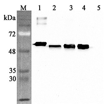
 were separated by 10% SDS-PAGE, and the membrane was blotted with Calreticulin antibody (GTX111627) diluted at 1:1000. The HRP-conjugated anti-rabbit IgG antibody (GTX213110-01) was used to detect the primary antibody.")
Calreticulin antibody detects Calreticulin protein at endoplasmic reticulum by immunofluorescent analysis. Sample: HeLa cells were fixed in ice-cold MeOH for 5 min. Green: Calreticulin stained by Calreticulin antibody (GTX111627) diluted at 1:500.
Calreticulin antibody
GTX111627
ApplicationsImmunoFluorescence, ImmunoPrecipitation, Western Blot, ImmunoCytoChemistry, ImmunoHistoChemistry, ImmunoHistoChemistry Paraffin
Product group Antibodies
ReactivityBovine, Human, Mouse, Rat
TargetCALR
Overview
- SupplierGeneTex
- Product NameCalreticulin antibody
- Delivery Days Customer9
- Application Supplier NoteWB: 1:500-1:3000. ICC/IF: 1:100-1:1000. IHC-P: 1:100-1:1000. *Optimal dilutions/concentrations should be determined by the researcher.Not tested in other applications.
- ApplicationsImmunoFluorescence, ImmunoPrecipitation, Western Blot, ImmunoCytoChemistry, ImmunoHistoChemistry, ImmunoHistoChemistry Paraffin
- CertificationResearch Use Only
- ClonalityPolyclonal
- Concentration0.64 mg/ml
- ConjugateUnconjugated
- Gene ID811
- Target nameCALR
- Target descriptioncalreticulin
- Target synonymsCALR1, CRT, HEL-S-99n, RO, SSA, cC1qR, calreticulin, CRP55, ERp60, HACBP, Sicca syndrome antigen A (autoantigen Ro; calreticulin), calregulin, endoplasmic reticulum resident protein 60, epididymis secretory sperm binding protein Li 99n, grp60
- HostRabbit
- IsotypeIgG
- Protein IDP27797
- Protein NameCalreticulin
- Scientific DescriptionCalreticulin is a multifunctional protein that acts as a major Ca(2+)-binding (storage) protein in the lumen of the endoplasmic reticulum. It is also found in the nucleus, suggesting that it may have a role in transcription regulation. Calreticulin binds to the synthetic peptide KLGFFKR, which is almost identical to an amino acid sequence in the DNA-binding domain of the superfamily of nuclear receptors. Calreticulin binds to antibodies in certain sera of systemic lupus and Sjogren patients which contain anti-Ro/SSA antibodies, it is highly conserved among species, and it is located in the endoplasmic and sarcoplasmic reticulum where it may bind calcium. The amino terminus of calreticulin interacts with the DNA-binding domain of the glucocorticoid receptor and prevents the receptor from binding to its specific glucocorticoid response element. Calreticulin can inhibit the binding of androgen receptor to its hormone-responsive DNA element and can inhibit androgen receptor and retinoic acid receptor transcriptional activities in vivo, as well as retinoic acid-induced neuronal differentiation. Thus, calreticulin can act as an important modulator of the regulation of gene transcription by nuclear hormone receptors. Systemic lupus erythematosus is associated with increased autoantibody titers against calreticulin but calreticulin is not a Ro/SS-A antigen. Earlier papers referred to calreticulin as an Ro/SS-A antigen but this was later disproven. Increased autoantibody titer against human calreticulin is found in infants with complete congenital heart block of both the IgG and IgM classes. [provided by RefSeq]
- ReactivityBovine, Human, Mouse, Rat
- Storage Instruction-20°C or -80°C,2°C to 8°C
- UNSPSC41116161
Datasheet
Related products
Product group Antibodies
CALR AntibodyCSB-PA001196
ApplicationsImmunoFluorescence, Western Blot, ELISA, ImmunoHistoChemistry
ReactivityHuman, Monkey, Mouse, Rat
TargetCALR
- SizePrice
Product group Antibodies
ApplicationsWestern Blot, ELISA
ReactivityHuman, Rat
- SizePrice

Product group Antibodies
Anti-Calreticulin/CALR Antibody Picoband(r)A00894-1-CARRIER-FREE
ApplicationsFlow Cytometry, Western Blot, ELISA, ImmunoHistoChemistry
ReactivityHuman, Mouse, Rat
TargetCALR
- SizePrice
Product group Antibodies

Anti-CALR [SAIC-16D-6B9]Ab00309-1.1
ApplicationsMass Spectrometry, Western Blot, ELISA
ReactivityHuman
TargetCALR
- SizePrice

Product group Antibodies
Anti-CALR AntibodyHPA002242
ApplicationsWestern Blot, ImmunoHistoChemistry
ReactivityHuman, Mouse, Rat
TargetCALR
- SizePrice
Product group Antibodies
References

Goat anti-calreticulinEB12387
ApplicationsWestern Blot, ELISA
ReactivityBovine, Canine, Human, Mouse, Porcine, Rat
TargetCALR
- SizePrice
Product group Antibodies

CALR / Calreticulin AntibodyLS-C400776
ApplicationsWestern Blot, ELISA, ImmunoHistoChemistry
ReactivityHuman, Mouse, Rat
TargetCALR
- SizePrice
Product group Antibodies
anti-Calreticulin (human), pAbAG-25A-0094
ApplicationsWestern Blot, ELISA
ReactivityHuman
TargetCALR
- SizePrice
Product group Antibodies
References
Calreticulin Polyclonal AntibodyBS-5913R
ApplicationsFlow Cytometry, ImmunoFluorescence, Western Blot, ELISA, ImmunoCytoChemistry, ImmunoHistoChemistry, ImmunoHistoChemistry Frozen, ImmunoHistoChemistry Paraffin
ReactivityHuman
TargetCALR
- SizePrice
Product group Antibodies
Calr Polyclonal AntibodyCAC07249
ApplicationsImmunoFluorescence, Western Blot, ELISA, ImmunoHistoChemistry
ReactivityMouse, Rat
TargetCALR
- SizePrice