

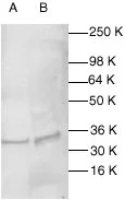
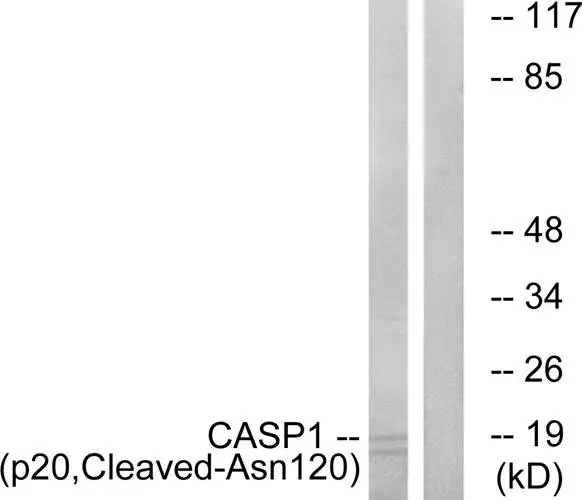
WB analysis of HeLa (A) and MCF-7 (B) whole cell lysates using GTX27978 Caspase 1 antibody. Dilution : 1:200
WB analysis of HeLa (A) and MCF-7 (B) whole cell lysates using GTX27978 Caspase 1 antibody. Dilution : 1:200
Caspase 1 antibody
GTX27978
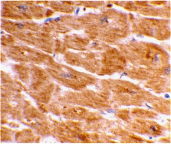
ApplicationsImmunoPrecipitation, Western Blot, ImmunoHistoChemistry
Product group Antibodies
ReactivityHuman
TargetCASP1
Overview
- SupplierGeneTex
- Product NameCaspase 1 antibody
- Delivery Days Customer9
- ApplicationsImmunoPrecipitation, Western Blot, ImmunoHistoChemistry
- CertificationResearch Use Only
- ClonalityPolyclonal
- Concentration0.2 mg/ml
- ConjugateUnconjugated
- Gene ID834
- Target nameCASP1
- Target descriptioncaspase 1
- Target synonymsICE, IL1BC, P45, caspase-1, IL-1 beta-converting enzyme, IL1B-convertase, caspase 1, apoptosis-related cysteine peptidase, interleukin 1, beta, convertase, interleukin 1-B converting enzyme
- HostRabbit
- IsotypeIgG
- Protein IDP29466
- Protein NameCaspase-1
- Scientific DescriptionThis gene encodes a protein which is a member of the cysteine-aspartic acid protease (caspase) family. Sequential activation of caspases plays a central role in the execution-phase of cell apoptosis. Caspases exist as inactive proenzymes which undergo proteolytic processing at conserved aspartic residues to produce 2 subunits, large and small, that dimerize to form the active enzyme. This gene was identified by its ability to proteolytically cleave and activate the inactive precursor of interleukin-1, a cytokine involved in the processes such as inflammation, septic shock, and wound healing. This gene has been shown to induce cell apoptosis and may function in various developmental stages. Studies of a similar gene in mouse suggest a role in the pathogenesis of Huntington disease. Alternative splicing results in transcript variants encoding distinct isoforms. [provided by RefSeq, Mar 2012]
- ReactivityHuman
- Storage Instruction2°C to 8°C
- UNSPSC12352203
Datasheet
Related products
Product group Antibodies
Casp1 Polyclonal AntibodyCAC10393
ApplicationsImmunoFluorescence, ELISA, ImmunoHistoChemistry
TargetCASP1
- SizePrice
Product group Antibodies
Anti-Casp3-1 (138-157) Antibody130-10600
ApplicationsELISA
ReactivityHuman
TargetCASP1
- SizePrice
Product group Antibodies
References
ApplicationsFlow Cytometry, ImmunoFluorescence, Western Blot, ELISA, ImmunoCytoChemistry, ImmunoHistoChemistry, ImmunoHistoChemistry Frozen, ImmunoHistoChemistry Paraffin
ReactivityHuman, Mouse, Rat
TargetCASP1
- SizePrice
Product group Antibodies
anti-Caspase-1 (p20) (human), mAb (Bally-1)AG-20B-0048
ApplicationsWestern Blot
ReactivityHuman
TargetCASP1
- SizePrice
Product group Antibodies
Anti-CASP1 AntibodyA99669
ApplicationsWestern Blot, ELISA
ReactivityHuman, Rat
- SizePrice
Product group Antibodies
ApplicationsWestern Blot
ReactivityHuman, Rat
TargetCASP1
- SizePrice
Product group Antibodies
Caspase 1 antibodyGTX31702
ApplicationsWestern Blot, ELISA, ImmunoHistoChemistry, ImmunoHistoChemistry Paraffin
ReactivityHuman
TargetCASP1
- SizePrice
Product group Antibodies
Caspase 1 antibody [HL2005]GTX637906
ApplicationsWestern Blot
ReactivityHuman
TargetCASP1
- SizePrice