

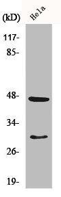
Whole cell extract (30 μg) was separated by 10% SDS-PAGE, and the membrane was blotted with Cathepsin D antibody (GTX133019) diluted at 1:500. The HRP-conjugated anti-rabbit IgG antibody (GTX213110-01) was used to detect the primary antibody.
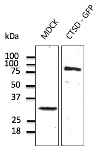
 were separated by 10% SDS-PAGE, and the membrane was blotted with Cathepsin D antibody (GTX133019) diluted at 1:1000. The HRP-conjugated anti-rabbit IgG antibody (GTX213110-01) was used to detect the primary antibody.")
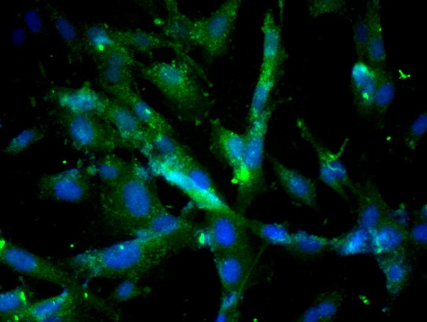
![Cathepsin D antibody detects Cathepsin D protein at lysosome by immunofluorescent analysis. Sample: MCF-7 cells were fixed in ice-cold MeOH for 5 min. Green: Cathepsin D stained by Cathepsin D antibody (GTX133019) diluted at 1:500. Red: alpha Tubulin, a cytoskeleton marker, stained by alpha Tubulin antibody [GT114] (GTX628802) diluted at 1:500. Blue: Hoechst 33342 staining.](https://www.genetex.com/upload/website/prouct_img/normal/GTX133019/GTX133019_42844_20180604_ICC_IF_w_23060523_313.webp "Cathepsin D antibody detects Cathepsin D protein at lysosome by immunofluorescent analysis. Sample: MCF-7 cells were fixed in ice-cold MeOH for 5 min. Green: Cathepsin D stained by Cathepsin D antibody (GTX133019) diluted at 1:500. Red: alpha Tubulin, a cytoskeleton marker, stained by alpha Tubulin antibody [GT114] (GTX628802) diluted at 1:500. Blue: Hoechst 33342 staining.")
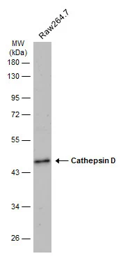
Whole cell extract (30 μg) was separated by 10% SDS-PAGE, and the membrane was blotted with Cathepsin D antibody (GTX133019) diluted at 1:500. The HRP-conjugated anti-rabbit IgG antibody (GTX213110-01) was used to detect the primary antibody.
Cathepsin D antibody
GTX133019
ApplicationsImmunoFluorescence, Western Blot, ImmunoCytoChemistry
Product group Antibodies
ReactivityHuman, Mouse
TargetCTSD
Overview
- SupplierGeneTex
- Product NameCathepsin D antibody
- Delivery Days Customer9
- Application Supplier NoteWB: 1:500-1:3000. ICC/IF: 1:100-1:1000. *Optimal dilutions/concentrations should be determined by the researcher.Not tested in other applications.
- ApplicationsImmunoFluorescence, Western Blot, ImmunoCytoChemistry
- CertificationResearch Use Only
- ClonalityPolyclonal
- Concentration0.56 mg/ml
- ConjugateUnconjugated
- Gene ID1509
- Target nameCTSD
- Target descriptioncathepsin D
- Target synonymsCLN10, CPSD, HEL-S-130P, cathepsin D, ceroid-lipofuscinosis, neuronal 10, epididymis secretory sperm binding protein Li 130P, lysosomal aspartyl peptidase, lysosomal aspartyl protease
- HostRabbit
- IsotypeIgG
- Protein IDP07339
- Protein NameCathepsin D
- Scientific DescriptionThis gene encodes a lysosomal aspartyl protease composed of a dimer of disulfide-linked heavy and light chains, both produced from a single protein precursor. This proteinase, which is a member of the peptidase C1 family, has a specificity similar to but narrower than that of pepsin A. Transcription of this gene is initiated from several sites, including one which is a start site for an estrogen-regulated transcript. Mutations in this gene are involved in the pathogenesis of several diseases, including breast cancer and possibly Alzheimer disease. [provided by RefSeq]
- ReactivityHuman, Mouse
- Storage Instruction-20°C or -80°C,2°C to 8°C
- UNSPSC41116161
Datasheet
Related products
Product group Antibodies
Anti-Cathepsin D Antibody128-10263
ApplicationsWestern Blot, ELISA
ReactivityHuman
- SizePrice
Product group Antibodies
Anti-Cathepsin D AntibodyA121574
ApplicationsImmunoFluorescence, Western Blot, ImmunoHistoChemistry, ImmunoHistoChemistry Frozen, ImmunoHistoChemistry Paraffin, Other Application
ReactivityCanine, Human, Monkey, Mouse, Rat
- SizePrice
Product group Antibodies
Cathepsin D Polyclonal AntibodyBS-1615R
ApplicationsImmunoFluorescence, Western Blot, ELISA, ImmunoCytoChemistry, ImmunoHistoChemistry, ImmunoHistoChemistry Frozen, ImmunoHistoChemistry Paraffin
ReactivityBovine, Canine, Human, Mouse, Porcine, Rabbit, Rat
TargetCTSD
- SizePrice
Product group Antibodies
CTSD AntibodyCSB-PA001329
ApplicationsWestern Blot, ELISA, ImmunoHistoChemistry
ReactivityHuman
TargetCTSD
- SizePrice
Product group Antibodies
CTSD Polyclonal AntibodyCAC13929
ApplicationsImmunoPrecipitation, ELISA, ImmunoHistoChemistry
TargetCTSD
- SizePrice
Product group Antibodies
Cathepsin D antibodyGTX20826
ApplicationsImmunoHistoChemistry, ImmunoHistoChemistry Frozen, ImmunoHistoChemistry Paraffin
ReactivityHuman
TargetCTSD
- SizePrice
Product group Antibodies
Cathepsin D antibody [CTD-19]GTX26313
ApplicationsWestern Blot, ELISA, ImmunoHistoChemistry, ImmunoHistoChemistry Paraffin
ReactivityHuman
TargetCTSD
- SizePrice
![IHC-P analysis of mouse heart tissue section using GTX01540 Cathepsin D antibody [GT1232]. Dilution : 1:100](https://www.genetex.com/upload/website/prouct_img/normal/GTX01540/GTX01540_20200508_IHC-P_w_23053121_156.webp)
Product group Antibodies
Cathepsin D antibody [GT1232]GTX01540
ApplicationsWestern Blot, ImmunoHistoChemistry, ImmunoHistoChemistry Paraffin
ReactivityHuman, Mouse
TargetCTSD
- SizePrice
Product group Antibodies

ApplicationsWestern Blot, ELISA
ReactivityHuman
TargetCTSD
- SizePrice