

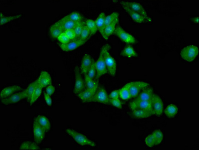
Immunofluorescence staining of HepG2 cells with CSB-PA019070LA01HU at 1:100, counter-stained with DAPI. The cells were fixed in 4% formaldehyde, permeabilized using 0.2% Triton X-100 and blocked in 10% normal Goat Serum. The cells were then incubated with the antibody overnight at 4°C. The secondary antibody was Alexa Fluor 488-congugated AffiniPure Goat Anti-Rabbit IgG(H+L).
Immunofluorescence staining of HepG2 cells with CSB-PA019070LA01HU at 1:100, counter-stained with DAPI. The cells were fixed in 4% formaldehyde, permeabilized using 0.2% Triton X-100 and blocked in 10% normal Goat Serum. The cells were then incubated with the antibody overnight at 4°C. The secondary antibody was Alexa Fluor 488-congugated AffiniPure Goat Anti-Rabbit IgG(H+L).
CAVIN1 Antibody
CSB-PA019070LA01HU
ApplicationsImmunoFluorescence, ELISA
Product group Antibodies
ReactivityHuman
TargetCAVIN1
Overview
- SupplierCusabio
- Product NameCAVIN1 Antibody
- Delivery Days Customer20
- ApplicationsImmunoFluorescence, ELISA
- CertificationResearch Use Only
- ClonalityPolyclonal
- ConjugateUnconjugated
- Gene ID284119
- Target nameCAVIN1
- Target descriptioncaveolae associated protein 1
- Target synonymsCAVIN, CGL4, FKSG13, PTRF, cavin-1, caveolae-associated protein 1, RNA polymerase I and transcript release factor, TTF-I interacting peptide 12, congenital generalized lipodystrophy 4, polymerase I and transcript release factor
- HostRabbit
- IsotypeIgG
- Protein IDQ6NZI2
- Protein NameCaveolae-associated protein 1
- Scientific DescriptionPlays an important role in caveolae formation and organization. Essential for the formation of caveolae in all tissues (PubMed:18056712, PubMed:18191225, PubMed:19726876). Core component of the CAVIN complex which is essential for recruitment of the complex to the caveolae in presence of calveolin-1 (CAV1). Essential for normal oligomerization of CAV1. Promotes ribosomal transcriptional activity in response to metabolic challenges in the adipocytes and plays an important role in the formation of the ribosomal transcriptional loop. Dissociates transcription complexes paused by DNA-bound TTF1, thereby releasing both RNA polymerase I and pre-RNA from the template (By similarity).
- ReactivityHuman
- Storage Instruction-20°C or -80°C
- UNSPSC41116161
Related products

Product group Antibodies
Anti-PTRF/CAVIN1 Antibody Picoband(r)A31732-2-CARRIER-FREE
ApplicationsFlow Cytometry, ImmunoFluorescence, Western Blot, ELISA, ImmunoCytoChemistry, ImmunoHistoChemistry
ReactivityHuman, Mouse, Rat
TargetCAVIN1
- SizePrice
Product group Antibodies
Anti-PTRF AntibodyA43775
ApplicationsWestern Blot
ReactivityHuman, Mouse, Rat
- SizePrice
Product group Antibodies

PTRF / CAVIN Antibody (aa150-200)LS-C288077
ApplicationsImmunoPrecipitation
ReactivityHuman
TargetCAVIN1
- SizePrice

Product group Antibodies
Anti-PTRF AntibodyHPA049838
ApplicationsWestern Blot, ImmunoCytoChemistry, ImmunoHistoChemistry
ReactivityHuman
TargetCAVIN1
- SizePrice