

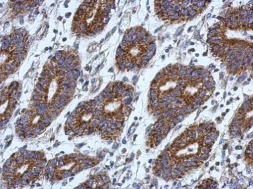
Immunohistochemical analysis of paraffin-embedded human colon carcinoma, using CCK4(GTX113816) antibody at 1:500 dilution.
Antigen Retrieval: Trilogy? (EDTA based, pH 8.0) buffer, 15min
![CCK4 antibody [N1N2], N-term detects CCK4 protein at cytoplasm by immunofluorescent analysis. Sample: HeLa cells were fixed in 4% paraformaldehyde at RT for 15 min. Green: CCK4 protein stained by CCK4 antibody [N1N2], N-term (GTX113816) diluted at 1:500. Blue: Hoechst 33342 staining. Scale bar = 10 μm.](https://www.genetex.com/upload/website/prouct_img/normal/GTX113816/GTX113816_40471_IFA_w_23060501_142.webp "CCK4 antibody [N1N2], N-term detects CCK4 protein at cytoplasm by immunofluorescent analysis. Sample: HeLa cells were fixed in 4% paraformaldehyde at RT for 15 min. Green: CCK4 protein stained by CCK4 antibody [N1N2], N-term (GTX113816) diluted at 1:500. Blue: Hoechst 33342 staining. Scale bar = 10 μm.")
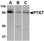
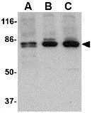
![Various whole cell extracts (30 μg) were separated by 7.5% SDS-PAGE, and the membrane was blotted with CCK4 antibody [N1N2], N-term (GTX113816) diluted at 1:1000. The HRP-conjugated anti-rabbit IgG antibody (GTX213110-01) was used to detect the primary antibody.](https://www.genetex.com/upload/website/prouct_img/normal/GTX113816/GTX113816_40471_20180126_WB_w_23060501_263.webp "Various whole cell extracts (30 μg) were separated by 7.5% SDS-PAGE, and the membrane was blotted with CCK4 antibody [N1N2], N-term (GTX113816) diluted at 1:1000. The HRP-conjugated anti-rabbit IgG antibody (GTX213110-01) was used to detect the primary antibody.")
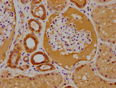
Immunohistochemical analysis of paraffin-embedded human colon carcinoma, using CCK4(GTX113816) antibody at 1:500 dilution.
Antigen Retrieval: Trilogy? (EDTA based, pH 8.0) buffer, 15min
CCK4 antibody [N1N2], N-term
GTX113816
ApplicationsImmunoFluorescence, Western Blot, ImmunoCytoChemistry, ImmunoHistoChemistry, ImmunoHistoChemistry Paraffin
Product group Antibodies
ReactivityHuman
TargetPTK7
Overview
- SupplierGeneTex
- Product NameCCK4 antibody [N1N2], N-term
- Delivery Days Customer9
- Application Supplier NoteWB: 1:500-1:3000. ICC/IF: 1:100-1:1000. IHC-P: 1:100-1:1000. *Optimal dilutions/concentrations should be determined by the researcher.Not tested in other applications.
- ApplicationsImmunoFluorescence, Western Blot, ImmunoCytoChemistry, ImmunoHistoChemistry, ImmunoHistoChemistry Paraffin
- CertificationResearch Use Only
- ClonalityPolyclonal
- Concentration1 mg/ml
- ConjugateUnconjugated
- Gene ID5754
- Target namePTK7
- Target descriptionprotein tyrosine kinase 7 (inactive)
- Target synonymsCCK-4, CCK4, inactive tyrosine-protein kinase 7, PTK7 protein tyrosine kinase 7, colon carcinoma kinase 4, pseudo tyrosine kinase receptor 7, tyrosine-protein kinase-like 7
- HostRabbit
- IsotypeIgG
- Protein IDQ13308
- Protein NameInactive tyrosine-protein kinase 7
- Scientific DescriptionReceptor protein tyrosine kinases transduce extracellular signals across the cell membrane. A subgroup of these kinases lack detectable catalytic tyrosine kinase activity but retain roles in signal transduction. The protein encoded by this gene is a member of this subgroup of tyrosine kinases and may function as a cell adhesion molecule. This gene is thought to be expressed in colon carcinomas but not in normal colon, and therefore may be a marker for or may be involved in tumor progression. Four transcript variants encoding four different isoforms have been found for this gene. [provided by RefSeq]
- ReactivityHuman
- Storage Instruction-20°C or -80°C,2°C to 8°C
- UNSPSC41116161
Datasheet
Related products
Product group Antibodies
PTK7 AntibodyCSB-PA018996LA01HU
ApplicationsELISA, ImmunoHistoChemistry
ReactivityHuman
TargetPTK7
- SizePrice
Product group Antibodies
Anti-PTK7 Antibody144-09839
ApplicationsWestern Blot, ImmunoHistoChemistry
ReactivityHuman, Mouse, Rat
TargetPTK7
- SizePrice

Product group Antibodies
ApplicationsFlow Cytometry, Western Blot, ImmunoHistoChemistry, ImmunoHistoChemistry Paraffin
ReactivityHuman
TargetPTK7
- SizePrice
Product group Antibodies
Anti-PTK7 AntibodyA48378
ApplicationsWestern Blot, ELISA, ImmunoHistoChemistry
ReactivityHuman, Mouse, Rat
- SizePrice

Product group Antibodies
Anti-PTK7 AntibodyHPA003222
ApplicationsWestern Blot, ImmunoHistoChemistry
ReactivityHuman
TargetPTK7
- SizePrice
Product group Antibodies

CCK4 / PTK7 AntibodyLS-C411361
ApplicationsWestern Blot, ImmunoHistoChemistry
ReactivityHuman, Mouse, Rat
TargetPTK7
- SizePrice
Product group Antibodies
CCK4 antibodyGTX31321
ApplicationsWestern Blot, ELISA, ImmunoHistoChemistry, ImmunoHistoChemistry Paraffin
ReactivityHuman, Mouse, Rat
TargetPTK7
- SizePrice
Product group Antibodies
CCK4 antibodyGTX31322
ApplicationsWestern Blot, ELISA, ImmunoHistoChemistry, ImmunoHistoChemistry Paraffin
ReactivityHuman, Mouse, Rat
TargetPTK7
- SizePrice