

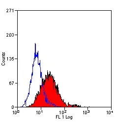
FACS analysis of KG1 cells using GTX11415 CD105 antibody [SN6] (FITC).
FACS analysis of KG1 cells using GTX11415 CD105 antibody [SN6] (FITC).
CD105 antibody [SN6] (FITC)
GTX11415
ApplicationsFlow Cytometry, ImmunoFluorescence, ImmunoCytoChemistry
Product group Antibodies
ReactivityEquine, Human, Monkey, Primate, Rabbit
TargetENG
Overview
- SupplierGeneTex
- Product NameCD105 antibody [SN6] (FITC)
- Delivery Days Customer9
- Application Supplier NoteFACS: Neat. *Optimal dilutions/concentrations should be determined by the researcher.Not tested in other applications.
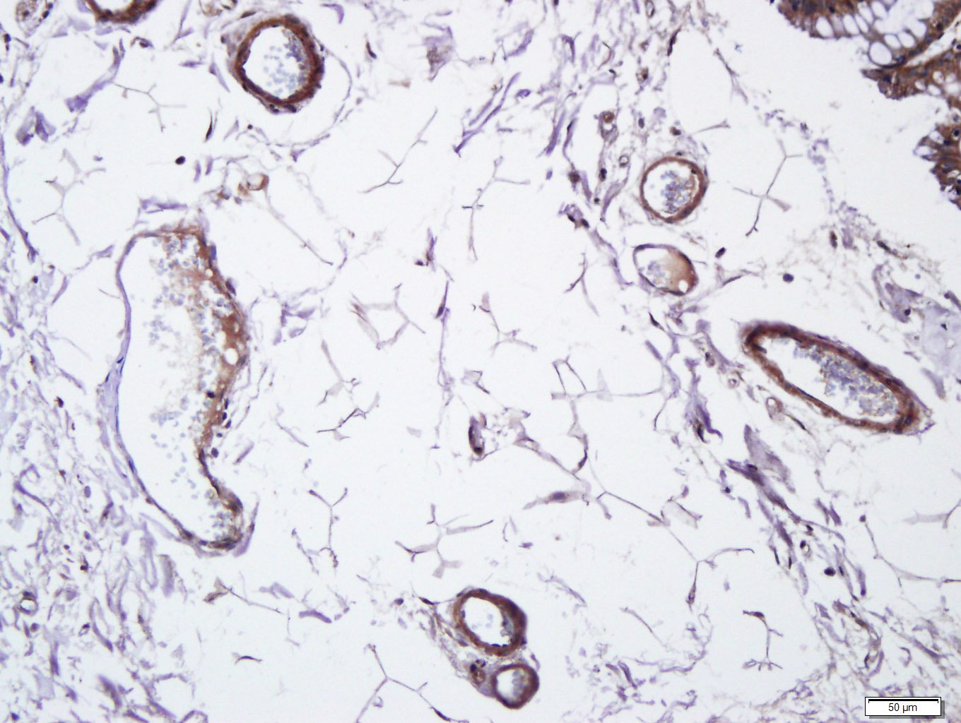
- ApplicationsFlow Cytometry, ImmunoFluorescence, ImmunoCytoChemistry
- CertificationResearch Use Only
- ClonalityMonoclonal
- Clone IDSN6
- Concentration0.1 mg/ml
- ConjugateFITC
- Gene ID2022
- Target nameENG
- Target descriptionendoglin
- Target synonymsEND, HHT1, ORW1, endoglin, CD105 antigen, soluble endoglin
- HostMouse
- IsotypeIgG1
- Protein IDP17813
- Protein NameEndoglin
- Scientific DescriptionThis gene encodes a homodimeric transmembrane protein which is a major glycoprotein of the vascular endothelium. This protein is a component of the transforming growth factor beta receptor complex and it binds to the beta1 and beta3 peptides with high affinity. Mutations in this gene cause hereditary hemorrhagic telangiectasia, also known as Osler-Rendu-Weber syndrome 1, an autosomal dominant multisystemic vascular dysplasia. This gene may also be involved in preeclampsia and several types of cancer. Alternatively spliced transcript variants encoding different isoforms have been found for this gene. [provided by RefSeq, May 2013]
- ReactivityEquine, Human, Monkey, Primate, Rabbit
- Storage Instruction-20°C,2°C to 8°C
- UNSPSC12352203
References
- Hosseinzadeh M, Kamali A, Hosseini S, et al. Higher Chondrogenic Potential of Extracellular Vesicles Derived from Mesenchymal Stem Cells Compared to Chondrocytes-EVs In Vitro. Biomed Res Int. 2021,2021:9011548. doi: 10.1155/2021/9011548Read this paper
- Li L, Chen Y, Fu Q, et al. Decellularized extracellular matrix loaded with IPFP-SC for repairing rabbit osteochondral defects. Am J Transl Res. 2021,13(10):11026-11047.Read this paper
- Wang Z, Zhang B, et al. Effect of Activated Platelet-Rich Plasma on Chondrogenic Differentiation of Rabbit Bone Marrow-Derived Mesenchymal Stem Cells. Stem Cells Int. 2021,2021:9947187. doi: 10.1155/2021/9947187Read this paper
- Tirpáková M, Vašíček J, Svoradová A, et al. Phenotypical Characterization and Neurogenic Differentiation of Rabbit Adipose Tissue-Derived Mesenchymal Stem Cells. Genes (Basel). 2021,12(3). doi: 10.3390/genes12030431Read this paper
- Li Q, Zhao F, Li Z, et al. Autologous Fractionated Adipose Tissue as a Natural Biomaterial and Novel One-Step Stem Cell Therapy for Repairing Articular Cartilage Defects. Front Cell Dev Biol. 2020,8:694. doi: 10.3389/fcell.2020.00694Read this paper
- Vašíček J, Kováč M, Baláži A, et al. Combined approach for characterization and quality assessment of rabbit bone marrow-derived mesenchymal stem cells intended for gene banking. N Biotechnol. 2020,54:1-12. doi: 10.1016/j.nbt.2019.08.001Read this paper
- Lin TK, Chen SD, Chuang YC, et al. Mitochondrial Transfer of Wharton's Jelly Mesenchymal Stem Cells Eliminates Mutation Burden and Rescues Mitochondrial Bioenergetics in Rotenone-Stressed MELAS Fibroblasts. Oxid Med Cell Longev. 2019,2019:9537504. doi: 10.1155/2019/9537504Read this paper
- Kulikova B, Kovac M, Bauer M, et al. Survivability of rabbit amniotic fluid-derived mesenchymal stem cells post slow-freezing or vitrification. Acta Histochem. 2019,121(4):491-499. doi: 10.1016/j.acthis.2019.03.008Read this paper
- Lee TC, Lee TH, Huang YH, et al. Comparison of surface markers between human and rabbit mesenchymal stem cells. PLoS One. 2014,9(11):e111390. doi: 10.1371/journal.pone.0111390Read this paper
Datasheet
Related products
Product group Antibodies
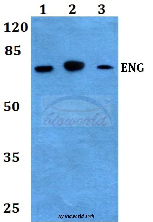
ApplicationsImmunoPrecipitation, Western Blot, ImmunoCytoChemistry, ImmunoHistoChemistry
TargetENG
- SizePrice
Product group Antibodies
Anti-Endoglin Antibody130-10018
ApplicationsELISA
ReactivityHuman
TargetENG
- SizePrice

Product group Antibodies
Anti-ENG AntibodyAMAB90925
ApplicationsImmunoHistoChemistry
ReactivityHuman
TargetENG
- SizePrice
Product group Antibodies
References
CD105 Polyclonal AntibodyBS-0579R
ApplicationsImmunoFluorescence, ImmunoHistoChemistry, ImmunoHistoChemistry Frozen, ImmunoHistoChemistry Paraffin
ReactivityMouse
TargetENG
- SizePrice
Product group Antibodies
Anti-Endoglin AntibodyA28219
ApplicationsWestern Blot
ReactivityHuman, Mouse
- SizePrice
Product group Antibodies
References
CD105 antibody [MJ7/18] (PE)GTX00572-08
ApplicationsFlow Cytometry
ReactivityHuman, Mouse, Rat
TargetENG
- SizePrice
Product group Antibodies
References
CD105 antibody [SN6] (PE)GTX75142
ApplicationsFlow Cytometry
ReactivityEquine, Human, Monkey, Primate, Rabbit
TargetENG
- SizePrice