

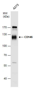
CD146 antibody detects CD146 protein by western blot analysis. Whole cell extracts (30 μg) was separated by 7.5 % SDS-PAGE, and blotted with CD146 antibody (GTX102413) diluted by 1:1000
![Wild-type (WT) and CD146 knockout (KO) HeLa cell extracts (30 μg) were separated by 5% SDS-PAGE, and the membrane was blotted with CD146 antibody [N1N3] (GTX102413) diluted at 1:500. The HRP-conjugated anti-rabbit IgG antibody (GTX213110-01) was used to detect the primary antibody.](https://www.genetex.com/upload/website/prouct_img/normal/GTX102413/GTX102413_40275_20181109_WB_KO_watermark_w_23060100_638.webp "Wild-type (WT) and CD146 knockout (KO) HeLa cell extracts (30 μg) were separated by 5% SDS-PAGE, and the membrane was blotted with CD146 antibody [N1N3] (GTX102413) diluted at 1:500. The HRP-conjugated anti-rabbit IgG antibody (GTX213110-01) was used to detect the primary antibody.")
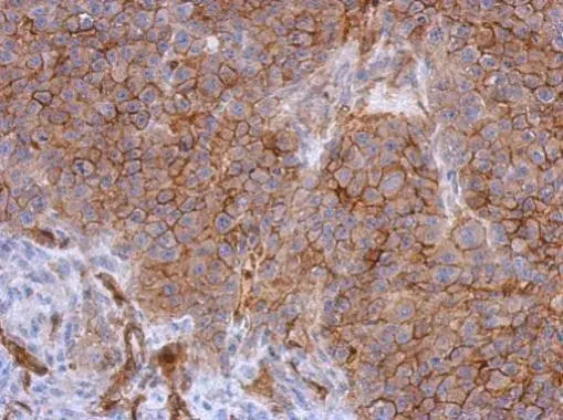
 antibody at 1:500 dilution.
Antigen Retrieval: Trilogy? (EDTA based, pH 8.0) buffer, 15min")
CD146 antibody detects CD146 protein by western blot analysis. Whole cell extracts (30 μg) was separated by 7.5 % SDS-PAGE, and blotted with CD146 antibody (GTX102413) diluted by 1:1000
CD146 antibody [N1N3]
GTX102413
ApplicationsWestern Blot, ImmunoHistoChemistry, ImmunoHistoChemistry Paraffin
Product group Antibodies
ReactivityHuman
TargetMCAM
Overview
- SupplierGeneTex
- Product NameCD146 antibody [N1N3]
- Delivery Days Customer9
- Application Supplier NoteWB: 1:500-1:3000. IHC-P: 1:100-1:1000. *Optimal dilutions/concentrations should be determined by the researcher.Not tested in other applications.
- ApplicationsWestern Blot, ImmunoHistoChemistry, ImmunoHistoChemistry Paraffin
- CertificationResearch Use Only
- ClonalityPolyclonal
- Concentration1 mg/ml
- ConjugateUnconjugated
- Gene ID4162
- Target nameMCAM
- Target descriptionmelanoma cell adhesion molecule
- Target synonymsCD146, HEMCAM, METCAM, MUC18, MelCAM, cell surface glycoprotein MUC18, Gicerin, S-endo 1 endothelial-associated antigen, cell surface glycoprotein P1H12, melanoma adhesion molecule, melanoma-associated antigen A32, melanoma-associated antigen MUC18
- HostRabbit
- IsotypeIgG
- Protein IDP43121
- Protein NameCell surface glycoprotein MUC18
- Scientific DescriptionPlays a role in cell adhesion, and in cohesion of the endothelial monolayer at intercellular junctions in vascular tissue. Its expression may allow melanoma cells to interact with cellular elements of the vascular system, thereby enhancing hematogeneous tumor spread. Could be an adhesion molecule active in neural crest cells during embryonic development. Acts as surface receptor that triggers tyrosine phosphorylation of FYN and PTK2, and a transient increase in the intracellular calcium concentration.
- ReactivityHuman
- Storage Instruction-20°C or -80°C,2°C to 8°C
- UNSPSC12352203
Datasheet
Related products
Product group Antibodies

Anti-CD146 [IR94]Ab00908-10.0
ApplicationsFlow Cytometry, Western Blot, ELISA, Neutralisation/Blocking
ReactivityHuman
TargetMCAM
- SizePrice
Product group Antibodies
Anti-MCAM Antibody144-60521
ApplicationsWestern Blot, ImmunoHistoChemistry
ReactivityHuman, Mouse, Rat
TargetMCAM
- SizePrice
Product group Antibodies

CD146 AntibodyABX242914
ApplicationsFlow Cytometry, ImmunoFluorescence, Western Blot, ELISA, ImmunoCytoChemistry, ImmunoHistoChemistry
- SizePrice
Product group Antibodies
References
CD146 antibody [C1C3]GTX108777
ApplicationsImmunoFluorescence, Western Blot, ImmunoCytoChemistry, ImmunoHistoChemistry, ImmunoHistoChemistry Paraffin
ReactivityHuman, Mouse, Rat
TargetMCAM
- SizePrice
![WB analysis of HEK293 (1) and CD146 (AA: 84-189)-hIgGFc transfected HEK293 (2) cell lysate using GTX60775 CD146 antibody [6C3E6].](https://www.genetex.com/upload/website/prouct_img/normal/GTX60775/GTX60775_20170912_WB_w_23061123_263.webp)
Product group Antibodies
References
CD146 antibody [6C3E6]GTX60775
ApplicationsFlow Cytometry, Western Blot, ELISA, ImmunoHistoChemistry, ImmunoHistoChemistry Paraffin
ReactivityHuman
TargetMCAM
- SizePrice
Product group Antibodies
Anti-MCAM AntibodyA30904
ApplicationsWestern Blot, ImmunoHistoChemistry
ReactivityHuman, Mouse, Rat
- SizePrice

Product group Antibodies
Anti-CD146/MCAM Antibody Picoband(r)RP1050-CARRIER-FREE
ApplicationsWestern Blot, ImmunoHistoChemistry
ReactivityHuman
TargetMCAM
- SizePrice
Product group Antibodies
CD146 Polyclonal AntibodyBS-1618R
ApplicationsFlow Cytometry, Western Blot, ELISA, ImmunoHistoChemistry, ImmunoHistoChemistry Paraffin
ReactivityBovine, Canine, Equine, Human, Mouse, Rat
TargetMCAM
- SizePrice