

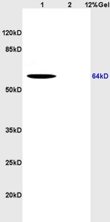
Lane 1: human colon carcinoma lysates Lane 2: rat brain lysates probed with Anti CD166 Polyclonal Antibody, Unconjugated (bs-1251R) at 1:200 in 4˚C. Followed by conjugation to secondary antibody (bs-0295G-HRP) at 1:3000 90min in 37˚C. Predicted band 64kD. Observed band size: 64kD.
Lane 1: human colon carcinoma lysates Lane 2: rat brain lysates probed with Anti CD166 Polyclonal Antibody, Unconjugated (bs-1251R) at 1:200 in 4˚C. Followed by conjugation to secondary antibody (bs-0295G-HRP) at 1:3000 90min in 37˚C. Predicted band 64kD. Observed band size: 64kD.
CD166 Polyclonal Antibody
BS-1251R
ApplicationsFlow Cytometry, ImmunoFluorescence, Western Blot, ELISA, ImmunoCytoChemistry, ImmunoHistoChemistry, ImmunoHistoChemistry Frozen, ImmunoHistoChemistry Paraffin
Product group Antibodies
ReactivityBovine, Canine, Chicken, Equine, Human, Mouse, Porcine, Rabbit, Rat
TargetALCAM
Overview
- SupplierBioss
- Product NameCD166 Polyclonal Antibody
- Delivery Days Customer16
- ApplicationsFlow Cytometry, ImmunoFluorescence, Western Blot, ELISA, ImmunoCytoChemistry, ImmunoHistoChemistry, ImmunoHistoChemistry Frozen, ImmunoHistoChemistry Paraffin
- Applications SupplierWB(1:300-5000), ELISA(1:500-1000), FCM(1:20-100), IHC-F(1:100-500), IF(IHC-P)(1:50-200), IF(IHC-F)(1:50-200), IF(ICC)(1:50-200)
- CertificationResearch Use Only
- ClonalityPolyclonal
- Concentration1 ug/ul
- ConjugateUnconjugated
- Gene ID214
- Target nameALCAM
- Target descriptionactivated leukocyte cell adhesion molecule
- Target synonymsCD166, MEMD, CD166 antigen
- HostRabbit
- IsotypeIgG
- Protein IDQ13740
- Protein NameCD166 antigen
- ReactivityBovine, Canine, Chicken, Equine, Human, Mouse, Porcine, Rabbit, Rat
- Storage Instruction-20°C
- UNSPSC41116161
References
- Combined approach for characterization and quality assessment of rabbit bone marrow-derived mesenchymal stem cells intended for gene banking. Vaicek J et al., 2020 Jan 25, N BiotechnolRead this paper
- Different RNA and protein expression of surface markers in rabbit amniotic fluid-derived mesenchymal stem cells. Kovac M et al., 2017 Nov, Biotechnol ProgRead this paper
Datasheet
Related products
Product group Antibodies
Anti-ALCAM AntibodyA97726
ApplicationsWestern Blot, ELISA
ReactivityHuman, Mouse, Rat
- SizePrice
Product group Antibodies

Anti-CD166 [EJ212/007-C12-5]Ab02669-10.0
ApplicationsFunctional Assay, ImmunoPrecipitation, Western Blot, ELISA, ImmunoHistoChemistry
ReactivityHuman
TargetALCAM
- SizePrice
Product group Antibodies
Anti-ALCAM Antibody144-65500
ApplicationsWestern Blot
ReactivityHuman, Mouse
TargetALCAM
- SizePrice

Product group Antibodies
Anti-CD166/ALCAM Antibody Picoband(r)A01788-1-CARRIER-FREE
ApplicationsFlow Cytometry, ImmunoFluorescence, Western Blot, ImmunoHistoChemistry
ReactivityHuman, Mouse, Rat
TargetALCAM
- SizePrice
Product group Antibodies
Alcam Polyclonal AntibodyCAC11068
ApplicationsImmunoFluorescence, ELISA, ImmunoHistoChemistry
TargetALCAM
- SizePrice
Product group Antibodies
ALCAM AntibodyCSB-PA005632
ApplicationsWestern Blot, ELISA
ReactivityHuman, Mouse, Rat
TargetALCAM
- SizePrice
Product group Antibodies

ALCAM / CD166 AntibodyLS-C408399
ApplicationsWestern Blot
ReactivityHuman
TargetALCAM
- SizePrice

Product group Antibodies
Anti-ALCAM AntibodyHPA010926
ApplicationsImmunoHistoChemistry
ReactivityHuman
TargetALCAM
- SizePrice
![CD166 antibody detects CD166 protein at cell membrane by immunohistochemical analysis. Sample: Paraffin-embedded mouse liver. Green: CD166 stained by CD166 antibody (GTX130098) diluted at 1:500. Red: beta Catenin, a cell membrane marker, stained by beta Catenin antibody [GT3171] (GTX632676) diluted at 1:500. Blue: Fluoroshield with DAPI (GTX30920). Antigen Retrieval: Citrate buffer, pH 6.0, 15 min](https://www.genetex.com/upload/website/prouct_img/normal/GTX130098/GTX130098_41948_20200214_IHC-P-FL_M_w_23060523_136.webp)
Product group Antibodies
CD166 antibodyGTX130098
ApplicationsWestern Blot, ImmunoHistoChemistry, ImmunoHistoChemistry Paraffin
ReactivityHuman, Mouse, Rat
TargetALCAM
- SizePrice