

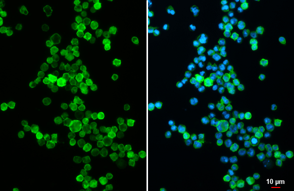
CD19 antibody [C1C3] detects CD19 protein at cell membrane by immunofluorescent analysis. Sample: Raji cells were fixed in 4% paraformaldehyde at RT for 15 min. Green: CD19 stained by CD19 antibody [C1C3] (GTX101512) diluted at 1:500. Blue: Fluoroshield with DAPI (GTX30920).
![Various whole cell extracts (30 μg) were separated by 7.5% SDS-PAGE, and the membrane was blotted with CD19 antibody [C1C3] (GTX101512) diluted at 1:1000. The HRP-conjugated anti-rabbit IgG antibody (GTX213110-01) was used to detect the primary antibody.](https://www.genetex.com/upload/website/prouct_img/normal/GTX101512/GTX101512_44090_20201009_WB_w_23060100_951.webp "Various whole cell extracts (30 μg) were separated by 7.5% SDS-PAGE, and the membrane was blotted with CD19 antibody [C1C3] (GTX101512) diluted at 1:1000. The HRP-conjugated anti-rabbit IgG antibody (GTX213110-01) was used to detect the primary antibody.")
![Mouse tissue extract (50 μg) was separated by 7.5% SDS-PAGE, and the membrane was blotted with CD19 antibody [C1C3] (GTX101512) diluted at 1:1000.](https://www.genetex.com/upload/website/prouct_img/normal/GTX101512/GTX101512_40422_20160204_WB_M_leukocyte_w_23060100_833.webp "Mouse tissue extract (50 μg) was separated by 7.5% SDS-PAGE, and the membrane was blotted with CD19 antibody [C1C3] (GTX101512) diluted at 1:1000.")
![CD19 antibody [C1C3] detects CD19 protein at cell membrane and cytoplasm by immunohistochemical analysis. Sample: Paraffin-embedded human tonsil. Green: CD19 stained by CD19 antibody [C1C3] (GTX101512) diluted at 1:250. Blue: Fluoroshield with DAPI (GTX30920). Antigen Retrieval: Citrate buffer, pH 6.0, 15 min](https://www.genetex.com/upload/website/prouct_img/normal/GTX101512/GTX101512_43397_20190531_IHC-P-FL_w_23060100_506.webp "CD19 antibody [C1C3] detects CD19 protein at cell membrane and cytoplasm by immunohistochemical analysis. Sample: Paraffin-embedded human tonsil. Green: CD19 stained by CD19 antibody [C1C3] (GTX101512) diluted at 1:250. Blue: Fluoroshield with DAPI (GTX30920). Antigen Retrieval: Citrate buffer, pH 6.0, 15 min")
![CD19 antibody [C1C3] (GTX101512) detects CD19 protein by flow cytometry analysis. Sample: mouse splenocytes cell fixed in 4% paraformaldehyde at 4oC for 15 min. Black: Unlabelled sample was used as a control. Red: CD19 antibody [C1C3] (GTX101512) dilution: 1:50. Acquisition of 20,000 events were collected for FACS analysis.](https://www.genetex.com/upload/website/prouct_img/normal/GTX101512/GTX101512_40422_20150609_FACS_w_23060100_523.webp "CD19 antibody [C1C3] (GTX101512) detects CD19 protein by flow cytometry analysis. Sample: mouse splenocytes cell fixed in 4% paraformaldehyde at 4oC for 15 min. Black: Unlabelled sample was used as a control. Red: CD19 antibody [C1C3] (GTX101512) dilution: 1:50. Acquisition of 20,000 events were collected for FACS analysis.")
![CD19 antibody [C1C3] detects CD19 protein at cell membrane by immunohistochemical analysis. Sample: Paraffin-embedded human tonsil. CD19 stained by CD19 antibody [C1C3] (GTX101512) diluted at 1:500. Antigen Retrieval: Citrate buffer, pH 6.0, 15 min](https://www.genetex.com/upload/website/prouct_img/normal/GTX101512/GTX101512_44090_20201120_IHC-P_w_23060100_526.webp "CD19 antibody [C1C3] detects CD19 protein at cell membrane by immunohistochemical analysis. Sample: Paraffin-embedded human tonsil. CD19 stained by CD19 antibody [C1C3] (GTX101512) diluted at 1:500. Antigen Retrieval: Citrate buffer, pH 6.0, 15 min")
CD19 antibody [C1C3] detects CD19 protein at cell membrane by immunofluorescent analysis. Sample: Raji cells were fixed in 4% paraformaldehyde at RT for 15 min. Green: CD19 stained by CD19 antibody [C1C3] (GTX101512) diluted at 1:500. Blue: Fluoroshield with DAPI (GTX30920).
CD19 antibody [C1C3]
GTX101512
ApplicationsFlow Cytometry, ImmunoFluorescence, Western Blot, ImmunoCytoChemistry, ImmunoHistoChemistry, ImmunoHistoChemistry Paraffin
Product group Antibodies
ReactivityHuman, Mouse
TargetCD19
Overview
- SupplierGeneTex
- Product NameCD19 antibody [C1C3]
- Delivery Days Customer9
- Application Supplier NoteWB: 1:500-1:3000. ICC/IF: 1:100-1:1000. IHC-P: 1:100-1:1000. FACS: 1:50-1:200. *Optimal dilutions/concentrations should be determined by the researcher.Not tested in other applications.
- ApplicationsFlow Cytometry, ImmunoFluorescence, Western Blot, ImmunoCytoChemistry, ImmunoHistoChemistry, ImmunoHistoChemistry Paraffin
- CertificationResearch Use Only
- ClonalityPolyclonal
- Concentration0.21 mg/ml
- ConjugateUnconjugated
- Gene ID930
- Target nameCD19
- Target descriptionCD19 molecule
- Target synonymsB4, CVID3, B-lymphocyte antigen CD19, B-lymphocyte surface antigen B4, T-cell surface antigen Leu-12, differentiation antigen CD19
- HostRabbit
- IsotypeIgG
- Protein IDP15391
- Protein NameB-lymphocyte antigen CD19
- Scientific DescriptionLymphocytes proliferate and differentiate in response to various concentrations of different antigens. The ability of the B cell to respond in a specific, yet sensitive manner to the various antigens is achieved with the use of low-affinity antigen receptors. This gene encodes a cell surface molecule which assembles with the antigen receptor of B lymphocytes in order to decrease the threshold for antigen receptor-dependent stimulation. [provided by RefSeq]
- ReactivityHuman, Mouse
- Storage Instruction-20°C or -80°C,2°C to 8°C
- UNSPSC12352203
References
- Yasuda T, Saito Y, Ono C, et al. Generation and characterization of CD19-iCre mice as a tool for efficient and specific conditional gene targeting in B cells. Sci Rep. 2021,11(1):5524. doi: 10.1038/s41598-021-84786-6Read this paper
- Kurimoto C, Inaba H, Ariyasu H, et al. Predictive and sensitive biomarkers for thyroid dysfunctions during treatment with immune-checkpoint inhibitors. Cancer Sci. 2020,111(5):1468-1477. doi: 10.1111/cas.14363Read this paper
- Bottero V, Sadagopan S, Johnson KE, et al. Kaposi's sarcoma-associated herpesvirus-positive primary effusion lymphoma tumor formation in NOD/SCID mice is inhibited by neomycin and neamine blocking angiogenin's nuclear translocation. J Virol. 2013,87(21):11806-20. doi: 10.1128/JVI.01920-13Read this paper
Datasheet
Related products
Product group Antibodies

Anti-CD19 [6D5]AB01489-2.0-VXS
ApplicationsFlow Cytometry, ImmunoPrecipitation, Western Blot, ImmunoHistoChemistry
ReactivityMouse
TargetCD19
- SizePrice
Product group Antibodies
ApplicationsFlow Cytometry
TargetCD19
- SizePrice
Product group Antibodies
Anti-CD19 Antibody144-02577
ApplicationsWestern Blot, ImmunoHistoChemistry
ReactivityHuman, Mouse, Rat
TargetCD19
- SizePrice

Product group Antibodies
Anti-CD19 Antibody Picoband(r)A00154-1-CARRIER-FREE
ApplicationsWestern Blot, ELISA
ReactivityHuman, Mouse
TargetCD19
- SizePrice
![FACS analysis of human peripheral blood lymphocytes using GTX01455-06 CD19 antibody [HIB19] (FITC). Solid lone : primary antibody Dashed line : isotype control antibody amount : 1 μg (5 μl)](https://www.genetex.com/upload/website/prouct_img/normal/GTX01455-06/GTX01455-06_20200428_FACS74_w_23053121_788.webp)
Product group Antibodies
References
CD19 antibody [HIB19] (FITC)GTX01455-06
ApplicationsFlow Cytometry
ReactivityHuman
TargetCD19
- SizePrice
![FACS analysis of human peripheral blood lymphocytes using GTX01455-07 CD19 antibody [HIB19] (APC). Solid lone : primary antibody Dashed line : isotype control antibody amount : 0.125 μg (5 μl)](https://www.genetex.com/upload/website/prouct_img/normal/GTX01455-07/GTX01455-07_20200428_FACS10_w_23053121_580.webp)
Product group Antibodies
References
CD19 antibody [HIB19] (APC)GTX01455-07
ApplicationsFlow Cytometry
ReactivityHuman
TargetCD19
- SizePrice
![FACS analysis of human peripheral blood lymphocytes using GTX01455-08 CD19 antibody [HIB19] (PE). Solid lone : primary antibody Dashed line : isotype control antibody amount : 0.25 μg (5 μl)](https://www.genetex.com/upload/website/prouct_img/normal/GTX01455-08/GTX01455-08_20200428_FACS100_w_23053121_847.webp)
Product group Antibodies
References
CD19 antibody [HIB19] (PE)GTX01455-08
ApplicationsFlow Cytometry
ReactivityHuman
TargetCD19
- SizePrice
![FACS analysis of human peripheral blood lymphocytes using GTX01455-09 CD19 antibody [HIB19] (PE-Cy5). Solid lone : primary antibody Dashed line : isotype control antibody amount : 0.25 μg (5 μl)](https://www.genetex.com/upload/website/prouct_img/normal/GTX01455-09/GTX01455-09_20200428_FACS133_w_23053121_861.webp)
Product group Antibodies
References
CD19 antibody [HIB19] (PE-Cy5)GTX01455-09
ApplicationsFlow Cytometry
ReactivityHuman
TargetCD19
- SizePrice
![FACS analysis of human peripheral blood lymphocytes using GTX01455-10 CD19 antibody [HIB19] (PE-Cy7). Solid lone : primary antibody Dashed line : isotype control antibody amount : 0.25 μg (5 μl)](https://www.genetex.com/upload/website/prouct_img/normal/GTX01455-10/GTX01455-10_20200428_FACS139_w_23053121_250.webp)
Product group Antibodies
References
CD19 antibody [HIB19] (PE-Cy7)GTX01455-10
ApplicationsFlow Cytometry
ReactivityHuman
TargetCD19
- SizePrice