

CD1D Antibody (clone 19G11)
LS-C810603
ApplicationsFlow Cytometry
Product group Antibodies
ReactivityMouse
TargetCD1D
Overview
- SupplierLifeSpan BioSciences
- Product NameCD1D Antibody (clone 19G11)
- Delivery Days Customer14
- Application Supplier NoteEach lot of this antibody is quality control tested by immunofluorescent staining with flow cytometric analysis. For flow cytometric staining, the suggested use of this reagent is = 0.5 microg per 106 cells in 100 microl volume or 100 microl of whole blood. It is recommended that the reagent be titrated for optimal performance for each application.. Flo Each lot of this antibody is quality control tested by immunofluorescent staining with flow cytometric analysis. For flow cytometric staining, the suggested use of this reagent is = 0.5 µg per 106 cells in 100 µl volume or 100 µl of whole blood. It is recommended that the reagent be titrated for optimal performance for each application.
- ApplicationsFlow Cytometry
- CertificationResearch Use Only
- ClonalityMonoclonal
- Clone ID19G11
- Concentration0.5 mg/ml
- ConjugateUnconjugated
- Gene ID912
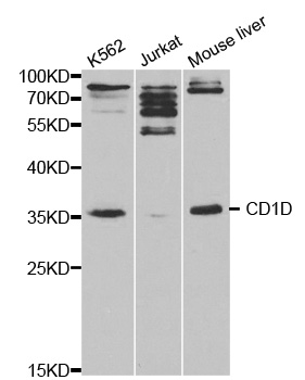
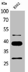
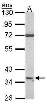
- Target nameCD1D
- Target descriptionCD1d molecule
- Target synonymsCD1A, R3, R3G1, antigen-presenting glycoprotein CD1d, CD1D antigen, d polypeptide, HMC class I antigen-like glycoprotein CD1D, T-cell surface glycoprotein CD1d, differentiation antigen CD1-alpha-3, thymocyte antigen CD1D
- HostRat
- IsotypeIgG2b
- ReactivityMouse
- Storage Instruction2°C to 8°C
- UNSPSC41116161
Related products
Product group Antibodies
Anti-CD1D AntibodyA29800
ApplicationsWestern Blot, ImmunoHistoChemistry
ReactivityHuman, Mouse, Rat
- SizePrice
Product group Antibodies
Anti-CD1D Antibody144-01760
ApplicationsImmunoFluorescence, Western Blot
ReactivityHuman, Mouse
TargetCD1D
- SizePrice
Product group Antibodies
CD1d Monoclonal AntibodyBSM-60537M
ApplicationsFlow Cytometry, ImmunoFluorescence, ImmunoCytoChemistry
ReactivityHuman
TargetCD1D
- SizePrice

Product group Antibodies
Anti-CD1D Antibody Picoband(r)A00323-CARRIER-FREE
ApplicationsFlow Cytometry, Western Blot, ELISA
ReactivityHuman, Rat
TargetCD1D
- SizePrice
Product group Antibodies
ApplicationsImmunoPrecipitation, Western Blot, ImmunoCytoChemistry, ImmunoHistoChemistry
ReactivityMouse, Porcine
TargetCD1D
- SizePrice
Product group Antibodies
CD1D AntibodyCSB-PA005192
ApplicationsWestern Blot, ELISA
ReactivityHuman
TargetCD1D
- SizePrice

Product group Antibodies
Anti-CD1D AntibodyHPA072662
ApplicationsImmunoCytoChemistry
ReactivityHuman
TargetCD1D
- SizePrice
Product group Antibodies
CD1d antibody [N2C3]GTX104898
ApplicationsWestern Blot
ReactivityHuman
TargetCD1D
- SizePrice