

![CD66 (CEA)(COL-1), CF405S conjugate, 0.1mg/mL [26628-22-8]](https://biotium.com/wp-content/uploads/2016/12/BNUB0002-1-1.jpg "CD66 (CEA)(COL-1), CF405S conjugate, 0.1mg/mL [26628-22-8]")
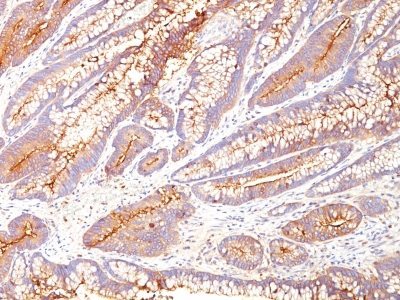
CD66 (CEA)(COL-1), CF405S conjugate, 0.1mg/mL [26628-22-8]
BNC040002
ApplicationsWestern Blot, ImmunoHistoChemistry, ImmunoHistoChemistry Paraffin
Product group Antibodies
ReactivityBovine, Human, Mouse
TargetCEACAM1
Overview
- SupplierBiotium
- Product NameCD66 (CEA)(COL-1), CF405S conjugate, 0.1mg/mL [26628-22-8]
- Delivery Days Customer9
- ApplicationsWestern Blot, ImmunoHistoChemistry, ImmunoHistoChemistry Paraffin
- CAS Number26628-22-8
- CertificationResearch Use Only
- ClonalityMonoclonal
- Clone IDCOL-1
- Concentration0.1 mg/ml
- ConjugateOther Conjugate
- Gene ID634
- Target nameCEACAM1
- Target descriptionCEA cell adhesion molecule 1
- Target synonymsBGP, BGP1, BGPI, cell adhesion molecule CEACAM1, CD66a antigen, antigen CD66, carcinoembryonic antigen related cell adhesion molecule 1, carcinoembryonic antigen-related cell adhesion molecule 1 (biliary glycoprotein)
- HostMouse
- IsotypeIgG2a
- Protein IDP06731
- Protein NameCell adhesion molecule CEACAM5
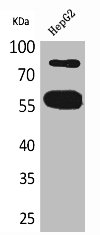
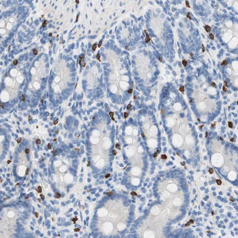
- Scientific DescriptionThis antibody recognizes proteins of 80-200 kDa, identified as different members of CEA family. CEA is synthesized during development in the fetal gut and is re-expressed in increased amounts in intestinal carcinomas and several other tumors. This MAb does not react with nonspecific cross-reacting antigen (NCA) and with human polymorphonuclear leucocytes. It shows no reaction with a variety of normal tissues and is suitable for staining of formalin/paraffin tissues. CEA is not found in benign glands, stroma, or malignant prostatic cells. Antibody to CEA is useful in detecting early foci of gastric carcinoma and in distinguishing pulmonary adenocarcinomas (60-70% are CEA positive) from pleural mesotheliomas (rarely or weakly CEA positive). Anti-CEA positivity is seen in adenocarcinomas from the lung, colon, stomach, esophagus, pancreas, gallbadder, urachus, salivary gland, ovary, and endocervix. Primary antibodies are available purified, or with a selection of fluorescent CF® Dyes and other labels. CF® Dyes offer exceptional brightness and photostability. Note: Conjugates of blue fluorescent dyes like CF®405S and CF®405M are not recommended for detecting low abundance targets, because blue dyes have lower fluorescence and can give higher non-specific background than other dye colors.
- SourceAnimal
- ReactivityBovine, Human, Mouse
- Storage Instruction2°C to 8°C,RT
- UNSPSC41116161
MSDS
Related products
Product group Antibodies
CEACAM1 AntibodyCSB-PA005134
ApplicationsWestern Blot, ELISA
ReactivityHuman
TargetCEACAM1
- SizePrice
Product group Antibodies
CEACAM1 Polyclonal AntibodyCAC14123
ApplicationsImmunoFluorescence, Western Blot, ELISA
TargetCEACAM1
- SizePrice
Product group Antibodies
Anti-CEACAM1 Antibody144-01702
ApplicationsWestern Blot
ReactivityHuman, Mouse, Rat
TargetCEACAM1
- SizePrice

Product group Antibodies
Anti-CEACAM1 Antibody Picoband(r)A00923-2-CARRIER-FREE
ApplicationsWestern Blot, ELISA
ReactivityHuman
TargetCEACAM1
- SizePrice
Product group Antibodies

Anti-CD66acd [YTH 71.3]Ab00198-23.0
ApplicationsFlow Cytometry, ImmunoHistoChemistry, ImmunoHistoChemistry Frozen
ReactivityHuman, Primate
TargetCEACAM1
- SizePrice
Product group Antibodies
Anti-CEACAM1 AntibodyA31625
ApplicationsWestern Blot, ImmunoHistoChemistry
ReactivityHuman, Mouse
- SizePrice
Product group Antibodies

CD66a / CEACAM1 AntibodyLS-C831499
ApplicationsImmunoHistoChemistry
ReactivityMouse, Rat
TargetCEACAM1
- SizePrice
Product group Antibodies
Anti-CEACAM1 AntibodyHPA011041
ApplicationsImmunoHistoChemistry
ReactivityHuman
TargetCEACAM1
- SizePrice