

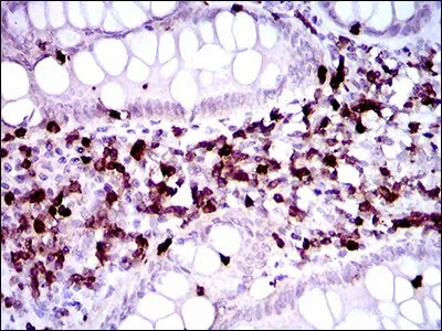
IHC-P analysis of endometrial cancer tissue using GTX60755 CD7 antibody [5B10E3].
![WB analysis of HEK293 (1) and CD7 (AA: 72-175)-hIgGFc transfected HEK293 (2) cell lysate using GTX60755 CD7 antibody [5B10E3].](https://www.genetex.com/upload/website/prouct_img/normal/GTX60755/GTX60755_20170912_WB_w_23061123_207.webp "WB analysis of HEK293 (1) and CD7 (AA: 72-175)-hIgGFc transfected HEK293 (2) cell lysate using GTX60755 CD7 antibody [5B10E3].")
![ELISA analysis of antigen using GTX60755 CD7 antibody [5B10E3].
Black : Control antigen 100ng
Purple : Antigen 10ng
Blue : Antigen 50ng
Red : Antigen 100ng](https://www.genetex.com/upload/website/prouct_img/normal/GTX60755/GTX60755_20170912_ELISA_w_23061123_716.webp "ELISA analysis of antigen using GTX60755 CD7 antibody [5B10E3].
Black : Control antigen 100ng
Purple : Antigen 10ng
Blue : Antigen 50ng
Red : Antigen 100ng")
IHC-P analysis of endometrial cancer tissue using GTX60755 CD7 antibody [5B10E3].
CD7 antibody [5B10E3]
GTX60755
ApplicationsFlow Cytometry, Western Blot, ELISA, ImmunoHistoChemistry, ImmunoHistoChemistry Paraffin
Product group Antibodies
ReactivityHuman
TargetCD7
Overview
- SupplierGeneTex
- Product NameCD7 antibody [5B10E3]
- Delivery Days Customer9
- Application Supplier NoteWB: 1/500 - 1/2000. IHC-P: 1/200 - 1/1000. FACS: 1/200 - 1/400. ELISA: 1/10000. *Optimal dilutions/concentrations should be determined by the researcher.Not tested in other applications.
- ApplicationsFlow Cytometry, Western Blot, ELISA, ImmunoHistoChemistry, ImmunoHistoChemistry Paraffin
- CertificationResearch Use Only
- ClonalityMonoclonal
- Clone ID5B10E3
- Concentration1 mg/ml
- ConjugateUnconjugated
- Gene ID924
- Target nameCD7
- Target descriptionCD7 molecule
- Target synonymsGP40, LEU-9, TP41, Tp40, T-cell antigen CD7, CD7 antigen (p41), T-cell leukemia antigen, T-cell surface antigen Leu-9, p41 protein
- HostMouse
- IsotypeIgG1
- Protein IDP09564
- Protein NameT-cell antigen CD7
- Scientific DescriptionThis gene encodes a transmembrane protein which is a member of the immunoglobulin superfamily. This protein is found on thymocytes and mature T cells. It plays an essential role in T-cell interactions and also in T-cell/B-cell interaction during early lymphoid development. [provided by RefSeq, Jul 2008]
- ReactivityHuman
- Storage Instruction-20°C or -80°C,2°C to 8°C
- UNSPSC12352203
Datasheet
Related products
Product group Antibodies

Anti-CD7 [3A1E]Ab00242-23.0
ApplicationsFunctional Assay, Flow Cytometry
ReactivityHuman
TargetCD7
- SizePrice

Product group Antibodies
Anti-CD7 Antibody Picoband(r)A01974-2-CARRIER-FREE
ApplicationsFlow Cytometry, Western Blot, ELISA, ImmunoCytoChemistry, ImmunoHistoChemistry
ReactivityHuman
TargetCD7
- SizePrice
![IHC-P analysis of human colorectal adenocarcinoma (COAD) tissue using GTX04390 CD7 antibody [MSVA-007R] HistoMAX?. Strong CD7 staining of a large fraction of tumor infiltrating lymphocytes in a colorectal adenocarcinoma. Tumor glands are completely CD7 negative.](https://www.genetex.com/upload/website/prouct_img/normal/GTX04390/GTX04390_20230728_IHC-P_14_23072722_645.webp)
Product group Antibodies
ApplicationsImmunoHistoChemistry, ImmunoHistoChemistry Paraffin
ReactivityHuman
TargetCD7
- SizePrice
![ELISA analysis of antigen using GTX60756 CD7 antibody [4D4F8].
Black : Control antigen 100ng
Purple : Antigen 10ng
Blue : Antigen 50ng
Red : Antigen 100ng](https://www.genetex.com/upload/website/prouct_img/normal/GTX60756/GTX60756_20170912_ELISA_w_23061123_131.webp)
Product group Antibodies
CD7 antibody [4D4F8]GTX60756
ApplicationsFlow Cytometry, Western Blot, ELISA, ImmunoHistoChemistry, ImmunoHistoChemistry Paraffin
ReactivityHuman
TargetCD7
- SizePrice
![FACS analysis of human peripheral blood using GTX79985 CD7 antibody [MEM-186] (APC).](https://www.genetex.com/upload/website/prouct_img/normal/GTX79985/GTX79985_20191025_AP_006_350_w_23061322_248.webp)
Product group Antibodies
CD7 antibody [MEM-186] (APC)GTX79985
ApplicationsFlow Cytometry
ReactivityHuman
TargetCD7
- SizePrice
![FACS analysis of human peripheral blood using GTX79986 CD7 antibody [MEM-186] (PE).](https://www.genetex.com/upload/website/prouct_img/normal/GTX79986/GTX79986_20191025_AP_006_351_w_23061322_781.webp)
Product group Antibodies
CD7 antibody [MEM-186] (PE)GTX79986
ApplicationsFlow Cytometry
ReactivityHuman
TargetCD7
- SizePrice
Product group Antibodies
Cd7 Polyclonal AntibodyCAC08296
ApplicationsELISA, ImmunoHistoChemistry
TargetCD7
- SizePrice