


![CD86(BU63), CF405S conjugate, 0.1mg/mL [26628-22-8]](https://biotium.com/wp-content/uploads/2016/12/BNUB0193-1-1.jpg "CD86(BU63), CF405S conjugate, 0.1mg/mL [26628-22-8]")
CD86(BU63), CF405S conjugate, 0.1mg/mL [26628-22-8]
BNC040193
ApplicationsFunctional Assay, Flow Cytometry, ImmunoFluorescence, ImmunoHistoChemistry, ImmunoHistoChemistry Paraffin
Product group Antibodies
ReactivityBovine, Human, Mouse, Rat
TargetCD86
Overview
- SupplierBiotium
- Product NameCD86(BU63), CF405S conjugate, 0.1mg/mL [26628-22-8]
- Delivery Days Customer9
- ApplicationsFunctional Assay, Flow Cytometry, ImmunoFluorescence, ImmunoHistoChemistry, ImmunoHistoChemistry Paraffin
- CAS Number26628-22-8
- CertificationResearch Use Only
- ClonalityMonoclonal
- Clone IDBU63
- Concentration0.1 mg/ml
- ConjugateOther Conjugate
- Gene ID942
- Target nameCD86
- Target descriptionCD86 molecule
- Target synonymsB7-2, B7.2, B70, CD28LG2, LAB72, T-lymphocyte activation antigen CD86, B-lymphocyte activation antigen B7-2, BU63, CD86 antigen (CD28 antigen ligand 2, B7-2 antigen), CD86 v6, CTLA-4 counter-receptor B7.2, FUN-1
- HostMouse
- IsotypeIgG1
- Protein IDP42081
- Protein NameT-lymphocyte activation antigen CD86
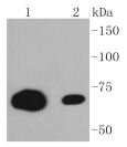
- Scientific DescriptionRecognizes a protein of 70 kDa, which is identified as CD86. CD86, along with CD80/B71, is an important accessory molecule in T cell co-stimulation via its interaction with CD28 and CD152/CTLA4, a cancer immunotherapy target. It is a type I transmembrane glycoprotein and a member of the immunoglobulin superfamily of cell surface receptors. It is expressed at high levels on resting peripheral monocytes and dendritic cells and at very low density on resting B and T lymphocytes. CD86 expression is rapidly upregulated by B cell specific stimuli with peak expression at 18 to 42 hours after stimulation. Since CD86 has rapid kinetics of induction, it is believed to be the major CD28 ligand expressed early in the immune response. It is also found on malignant Hodgkin and Reed Sternberg (HRS) cells in Hodgkins disease. Primary antibodies are available purified, or with a selection of fluorescent CF® Dyes and other labels. CF® Dyes offer exceptional brightness and photostability. Note: Conjugates of blue fluorescent dyes like CF®405S and CF®405M are not recommended for detecting low abundance targets, because blue dyes have lower fluorescence and can give higher non-specific background than other dye colors.
- SourceAnimal
- ReactivityBovine, Human, Mouse, Rat
- Storage Instruction2°C to 8°C,RT
- UNSPSC41116161
MSDS
Related products
Product group Antibodies
CD86 AntibodyCSB-PA004965EA01HU
ApplicationsELISA
ReactivityHuman
TargetCD86
- SizePrice

Product group Antibodies
Anti-CD86 Antibody Picoband(r)A00220-3-CARRIER-FREE
ApplicationsFlow Cytometry, ImmunoFluorescence, Western Blot, ELISA, ImmunoCytoChemistry
ReactivityHuman
TargetCD86
- SizePrice
Product group Antibodies
ApplicationsFlow Cytometry, ImmunoPrecipitation, Western Blot
ReactivityHuman
TargetCD86
- SizePrice
Product group Antibodies

Anti-CD86 [BU63]Ab00859-1.1
ApplicationsFlow Cytometry, ImmunoPrecipitation, Western Blot, ImmunoHistoChemistry
ReactivityHuman
TargetCD86
- SizePrice
Product group Antibodies
Anti-CD86 AntibodyA32352
ApplicationsFlow Cytometry, ImmunoFluorescence, Western Blot, ImmunoCytoChemistry, ImmunoHistoChemistry
ReactivityHuman, Monkey, Rat
- SizePrice
Product group Antibodies

CD86 Antibody (clone GL-1)LS-C770058
ApplicationsFlow Cytometry, ImmunoFluorescence, ImmunoPrecipitation, ImmunoHistoChemistry
ReactivityMouse
TargetCD86
- SizePrice

Product group Antibodies
Anti-CD86 AntibodyHPA026802
ApplicationsImmunoCytoChemistry
ReactivityHuman
TargetCD86
- SizePrice

Product group Antibodies
CD86 Recombinant AntibodyBSM-52375R
ApplicationsWestern Blot
ReactivityHuman, Mouse, Rat
TargetCD86
- SizePrice
Product group Antibodies
Anti-Human CD86 Antibody, PE150-10519
ReactivityHuman, Monkey, Primate
TargetCD86
- SizePrice