

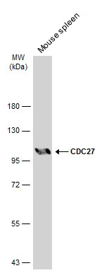
Mouse tissue extract (50 μg) was separated by 7.5% SDS-PAGE, and the membrane was blotted with CDC27 antibody (GTX111249) diluted at 1:500. The HRP-conjugated anti-rabbit IgG antibody (GTX213110-01) was used to detect the primary antibody, and the signal was developed with Trident ECL plus-Enhanced.
 were separated by 7.5% SDS-PAGE, and the membrane was blotted with CDC27 antibody (GTX111249) diluted at 1:500. The HRP-conjugated anti-rabbit IgG antibody (GTX213110-01) was used to detect the primary antibody, and the signal was developed with Trident ECL plus-Enhanced.")
Mouse tissue extract (50 μg) was separated by 7.5% SDS-PAGE, and the membrane was blotted with CDC27 antibody (GTX111249) diluted at 1:500. The HRP-conjugated anti-rabbit IgG antibody (GTX213110-01) was used to detect the primary antibody, and the signal was developed with Trident ECL plus-Enhanced.
CDC27 antibody
GTX111249
ApplicationsWestern Blot
Product group Antibodies
ReactivityHuman, Mouse
TargetCDC27
Overview
- SupplierGeneTex
- Product NameCDC27 antibody
- Delivery Days Customer9
- Application Supplier NoteWB: 1:500-1:3000. *Optimal dilutions/concentrations should be determined by the researcher.Not tested in other applications.
- ApplicationsWestern Blot
- CertificationResearch Use Only
- ClonalityPolyclonal
- Concentration1 mg/ml
- ConjugateUnconjugated
- Gene ID996
- Target nameCDC27
- Target descriptioncell division cycle 27
- Target synonymsANAPC3, APC3, CDC27Hs, D0S1430E, D17S978E, H-NUC, HNUC, NUC2, cell division cycle protein 27 homolog, D0S1430E, D17S978E, anaphase promoting complex subunit 3, anaphase-promoting complex, protein 3, cell division cycle 27 homolog, nuc2 homolog
- HostRabbit
- IsotypeIgG
- Protein IDP30260
- Protein NameCell division cycle protein 27 homolog
- Scientific DescriptionThe protein encoded by this gene shares strong similarity with Saccharomyces cerevisiae protein Cdc27, and the gene product of Schizosaccharomyces pombe nuc 2. This protein is a component of anaphase-promoting complex (APC), which is composed of eight protein subunits and highly conserved in eucaryotic cells. APC catalyzes the formation of cyclin B-ubiquitin conjugate that is responsible for the ubiquitin-mediated proteolysis of B-type cyclins. This protein and 3 other members of the APC complex contain the TPR (tetratricopeptide repeat), a protein domain important for protein-protein interaction. This protein was shown to interact with mitotic checkpoint proteins including Mad2, p55CDC and BUBR1, and thus may be involved in controlling the timing of mitosis. Alternatively spliced transcript variants encoding different isoforms have been found for this gene. [provided by RefSeq]
- ReactivityHuman, Mouse
- Storage Instruction-20°C or -80°C,2°C to 8°C
- UNSPSC41116161
Datasheet
Related products
Product group Antibodies
Anti-Cdc27 AntibodyA11494
ApplicationsImmunoFluorescence, Western Blot, ImmunoCytoChemistry, ImmunoHistoChemistry
ReactivityHuman, Mouse, Rat
- SizePrice

Product group Antibodies
Anti-CDC27 Antibody Picoband(r)A03905-3-CARRIER-FREE
ApplicationsFlow Cytometry, ImmunoFluorescence, Western Blot, ELISA, ImmunoCytoChemistry, ImmunoHistoChemistry
ReactivityHuman, Mouse, Rat
TargetCDC27
- SizePrice
Product group Antibodies
CDC27 Recombinant Antibody, Biotin ConjugatedBSM-61336R-BIOTIN
ApplicationsWestern Blot
TargetCDC27
- SizePrice
Product group Antibodies
CDC27 AntibodyCSB-PA001504
ApplicationsWestern Blot, ELISA, ImmunoHistoChemistry
ReactivityHuman, Mouse, Rat
TargetCDC27
- SizePrice
Product group Antibodies

CDC27 AntibodyLS-C403904
ApplicationsELISA, ImmunoHistoChemistry
ReactivityHuman, Mouse, Rat
TargetCDC27
- SizePrice
Product group Antibodies
CDC27 antibodyGTX17058
ApplicationsWestern Blot, ELISA, ImmunoHistoChemistry, ImmunoHistoChemistry Paraffin
ReactivityHuman, Mouse, Rat
TargetCDC27
- SizePrice
![IHC-P analysis of lung cancer tissue (left) and colon cancer tissue (right) using GTX82818 CDC27 antibody [5C12].](https://www.genetex.com/upload/website/prouct_img/normal/GTX82818/GTX82818_20170912_IHC-P_w_23051501_413.webp)
Product group Antibodies
CDC27 antibody [5C12]GTX82818
ApplicationsWestern Blot, ELISA, ImmunoHistoChemistry, ImmunoHistoChemistry Paraffin
ReactivityHuman
TargetCDC27
- SizePrice
Product group Antibodies
Anti-CDC27 AntibodyHPA028129
ApplicationsWestern Blot, ImmunoCytoChemistry, ImmunoHistoChemistry
ReactivityHuman
TargetCDC27
- SizePrice
![Western blot using Affinity Purified anti-cdc27 antibody (GTX41386) shows detection of a band ~90 kDa corresponding to human cdc27 (arrowhead). Approximately 35 ug of HeLa whole cell lysate was separated by SDS-PAGE and transferred onto nitrocellulose. After blocking the membrane was probed with the primary antibody diluted to 1.0 ug/ml for 2 h at room temperature followed by washes and reaction with a 1:10,000 dilution of IRDye800 conjugated Gt-a-Rabbit IgG [H&L] MX for 45 min at room temperature. IRDye800 fluorescence image was captured using the OdysseyR Infrared Imaging System developed by LI-COR.](https://www.genetex.com/upload/website/prouct_img/normal/GTX41386/GTX41386_20160330_WB_w_23060820_577.webp)
Product group Antibodies
CDC27 antibodyGTX41386
ApplicationsImmunoFluorescence, Western Blot, ELISA, ImmunoCytoChemistry
ReactivityHuman
TargetCDC27
- SizePrice