


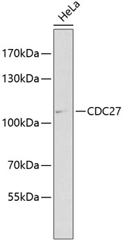
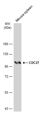
Western blot using Affinity Purified anti-cdc27 antibody (GTX41386) shows detection of a band ~90 kDa corresponding to human cdc27 (arrowhead). Approximately 35 ug of HeLa whole cell lysate was separated by SDS-PAGE and transferred onto nitrocellulose. After blocking the membrane was probed with the primary antibody diluted to 1.0 ug/ml for 2 h at room temperature followed by washes and reaction with a 1:10,000 dilution of IRDye800 conjugated Gt-a-Rabbit IgG [H&L] MX for 45 min at room temperature. IRDye800 fluorescence image was captured using the OdysseyR Infrared Imaging System developed by LI-COR.
Western blot using Affinity Purified anti-cdc27 antibody (GTX41386) shows detection of a band ~90 kDa corresponding to human cdc27 (arrowhead). Approximately 35 ug of HeLa whole cell lysate was separated by SDS-PAGE and transferred onto nitrocellulose. After blocking the membrane was probed with the primary antibody diluted to 1.0 ug/ml for 2 h at room temperature followed by washes and reaction with a 1:10,000 dilution of IRDye800 conjugated Gt-a-Rabbit IgG [H&L] MX for 45 min at room temperature. IRDye800 fluorescence image was captured using the OdysseyR Infrared Imaging System developed by LI-COR.
CDC27 antibody
GTX41386
ApplicationsImmunoFluorescence, Western Blot, ELISA, ImmunoCytoChemistry
Product group Antibodies
ReactivityHuman
TargetCDC27
Overview
- SupplierGeneTex
- Product NameCDC27 antibody
- Delivery Days Customer9
- Application Supplier NoteWB: 1:500-1:2000. ELISA: 1:5000-1:40000. *Optimal dilutions/concentrations should be determined by the researcher.Not tested in other applications.
- ApplicationsImmunoFluorescence, Western Blot, ELISA, ImmunoCytoChemistry
- CertificationResearch Use Only
- ClonalityPolyclonal
- Concentration1 mg/ml
- ConjugateUnconjugated
- Gene ID996
- Target nameCDC27
- Target descriptioncell division cycle 27
- Target synonymsANAPC3, APC3, CDC27Hs, D0S1430E, D17S978E, H-NUC, HNUC, NUC2, cell division cycle protein 27 homolog, D0S1430E, D17S978E, anaphase promoting complex subunit 3, anaphase-promoting complex, protein 3, cell division cycle 27 homolog, nuc2 homolog
- HostRabbit
- IsotypeIgG
- Protein IDP30260
- Protein NameCell division cycle protein 27 homolog
- Scientific DescriptionCdc27 shares strong similarity with Saccharomyces cerevisiae protein Cdc27, and the gene product of Schizosaccharomyces pombe nuc 2. It is a component of the Anaphase Promoting Complex (APC), which is composed of eight protein subunits and is highly conserved in eucaryotic cells. The APC catalyzes the formation of the cyclin B-ubiquitin conjugate that is responsible for the ubiquitin-mediated proteolysis of B-type cyclins. This protein and 3 other members of the APC complex contain the TPR (tetratricopeptide repeat), a protein domain important for protein-protein interaction. This protein was shown to interact with mitotic checkpoint proteins including Mad2, p55CDC and BUBR1, and thus may be involved in controlling the timing of mitosis.
- ReactivityHuman
- Storage Instruction-20°C or -80°C,2°C to 8°C
- UNSPSC41116161
Datasheet
Related products
Product group Antibodies
Anti-Cdc27 AntibodyA11494
ApplicationsImmunoFluorescence, Western Blot, ImmunoCytoChemistry, ImmunoHistoChemistry
ReactivityHuman, Mouse, Rat
- SizePrice

Product group Antibodies
Anti-CDC27 Antibody Picoband(r)A03905-3-CARRIER-FREE
ApplicationsFlow Cytometry, ImmunoFluorescence, Western Blot, ELISA, ImmunoCytoChemistry, ImmunoHistoChemistry
ReactivityHuman, Mouse, Rat
TargetCDC27
- SizePrice
Product group Antibodies
CDC27 Recombinant Antibody, Biotin ConjugatedBSM-61336R-BIOTIN
ApplicationsWestern Blot
TargetCDC27
- SizePrice
Product group Antibodies
CDC27 AntibodyCSB-PA001504
ApplicationsWestern Blot, ELISA, ImmunoHistoChemistry
ReactivityHuman, Mouse, Rat
TargetCDC27
- SizePrice
Product group Antibodies

CDC27 AntibodyLS-C403904
ApplicationsELISA, ImmunoHistoChemistry
ReactivityHuman, Mouse, Rat
TargetCDC27
- SizePrice
Product group Antibodies
CDC27 antibodyGTX17058
ApplicationsWestern Blot, ELISA, ImmunoHistoChemistry, ImmunoHistoChemistry Paraffin
ReactivityHuman, Mouse, Rat
TargetCDC27
- SizePrice
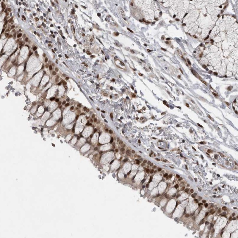
![IHC-P analysis of lung cancer tissue (left) and colon cancer tissue (right) using GTX82818 CDC27 antibody [5C12].](https://www.genetex.com/upload/website/prouct_img/normal/GTX82818/GTX82818_20170912_IHC-P_w_23051501_413.webp)
Product group Antibodies
CDC27 antibody [5C12]GTX82818
ApplicationsWestern Blot, ELISA, ImmunoHistoChemistry, ImmunoHistoChemistry Paraffin
ReactivityHuman
TargetCDC27
- SizePrice
Product group Antibodies
Anti-CDC27 AntibodyHPA028129
ApplicationsWestern Blot, ImmunoCytoChemistry, ImmunoHistoChemistry
ReactivityHuman
TargetCDC27
- SizePrice
Product group Antibodies
CDC27 antibodyGTX111249
ApplicationsWestern Blot
ReactivityHuman, Mouse
TargetCDC27
- SizePrice