

CDCP1 antibody
10R-3641
Product group Antibodies
Overview
- SupplierBiosynth
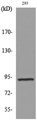
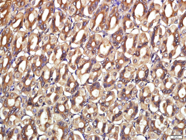
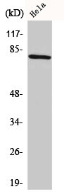
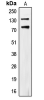
- Product NameCDCP1 antibody
- Delivery Days Customer11
- CertificationResearch Use Only
- UNSPSC41116161
Related products
Product group Antibodies
Anti-CDCP1 AntibodyA97649
ApplicationsWestern Blot, ELISA
ReactivityHuman, Mouse, Rat
- SizePrice

Product group Antibodies
Anti-CDCP1 Antibody Picoband(r)A02411-1-CARRIER-FREE
ApplicationsWestern Blot, ELISA
ReactivityHuman
TargetCDCP1
- SizePrice
Product group Antibodies
Anti-CDCP1 Antibody144-08254
ApplicationsWestern Blot
ReactivityHuman, Mouse
TargetCDCP1
- SizePrice
Product group Antibodies
CDCP1 Polyclonal AntibodyBS-5880R
ApplicationsImmunoFluorescence, Western Blot, ELISA, ImmunoCytoChemistry, ImmunoHistoChemistry, ImmunoHistoChemistry Frozen, ImmunoHistoChemistry Paraffin
ReactivityChicken, Human, Mouse, Rat
TargetCDCP1
- SizePrice
Product group Antibodies
CDCP1 AntibodyCSB-PA001521
ApplicationsWestern Blot, ELISA
ReactivityHuman, Mouse
TargetCDCP1
- SizePrice
Product group Antibodies

ApplicationsWestern Blot, ELISA, ImmunoHistoChemistry
ReactivityHuman
TargetCDCP1
- SizePrice
Product group Antibodies
ApplicationsImmunoPrecipitation, Western Blot, ImmunoCytoChemistry, ImmunoHistoChemistry
TargetCDCP1
- SizePrice
Product group Antibodies

CDCP1 AntibodyLS-C403055
ApplicationsWestern Blot, ELISA, ImmunoHistoChemistry
ReactivityHuman
TargetCDCP1
- SizePrice

Product group Antibodies
Anti-CDCP1 AntibodyHPA010978
ApplicationsImmunoHistoChemistry
ReactivityHuman
TargetCDCP1
- SizePrice
Product group Antibodies
CDCP1 (phospho Tyr707) antibodyGTX54967
ApplicationsWestern Blot
ReactivityHuman
TargetCDCP1
- SizePrice