

_w_23053123_883.webp)
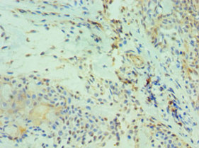
IHC-P analysis of human esophageal tissue section using GTX02852 CDH13 antibody [GT1255]. Dilution : 1:100
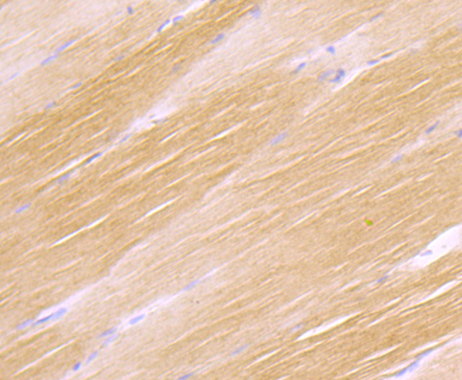
![IHC-P analysis of rat brain tissue section using GTX02852 CDH13 antibody [GT1255]. Dilution : 1:100](https://www.genetex.com/upload/website/prouct_img/normal/GTX02852/A3244_IHC_01_(1073418)_w_23053123_331.webp "IHC-P analysis of rat brain tissue section using GTX02852 CDH13 antibody [GT1255]. Dilution : 1:100")
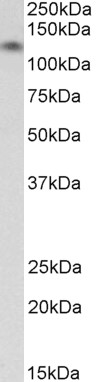
![WB analysis of mouse heart tissue lysates using GTX02852 CDH13 antibody [GT1255]. Dilution : 1:1000 Loading : 25μg](https://www.genetex.com/upload/website/prouct_img/normal/GTX02852/CutImage_A3244_WB_01_(1073926)_w_23053123_429.webp "WB analysis of mouse heart tissue lysates using GTX02852 CDH13 antibody [GT1255]. Dilution : 1:1000 Loading : 25μg")
![Whole cell extract (30 μg) was separated by 5% SDS-PAGE, and the membrane was blotted with CDH13 antibody [GT1255] (GTX02852) diluted at 1:500. The HRP-conjugated anti-rabbit IgG antibody (GTX213110-01) was used to detect the primary antibody.](https://www.genetex.com/upload/website/prouct_img/normal/GTX02852/GTX02852_4000001931_20210122_WB_2_w_23053123_929.webp "Whole cell extract (30 μg) was separated by 5% SDS-PAGE, and the membrane was blotted with CDH13 antibody [GT1255] (GTX02852) diluted at 1:500. The HRP-conjugated anti-rabbit IgG antibody (GTX213110-01) was used to detect the primary antibody.")
![Whole cell extract (30 μg) was separated by 5% SDS-PAGE, and the membrane was blotted with CDH13 antibody [GT1255] (GTX02852) diluted at 1:1000. The HRP-conjugated anti-rabbit IgG antibody (GTX213110-01) was used to detect the primary antibody, and the signal was developed with Trident ECL plus-Enhanced.](https://www.genetex.com/upload/website/prouct_img/normal/GTX02852/GTX02852_4000001931_20210122_WB_w_23053123_521.webp "Whole cell extract (30 μg) was separated by 5% SDS-PAGE, and the membrane was blotted with CDH13 antibody [GT1255] (GTX02852) diluted at 1:1000. The HRP-conjugated anti-rabbit IgG antibody (GTX213110-01) was used to detect the primary antibody, and the signal was developed with Trident ECL plus-Enhanced.")
![IHC-P analysis of mouse kidney tissue section using GTX02852 CDH13 antibody [GT1255]. Dilution : 1:100](https://www.genetex.com/upload/website/prouct_img/normal/GTX02852/A3244_IHC_03_(1073420)_w_23053123_211.webp "IHC-P analysis of mouse kidney tissue section using GTX02852 CDH13 antibody [GT1255]. Dilution : 1:100")
![Mouse tissue extract (50 μg) was separated by 5% SDS-PAGE, and the membrane was blotted with CDH13 antibody [GT1255] (GTX02852) diluted at 1:1000. The HRP-conjugated anti-rabbit IgG antibody (GTX213110-01) was used to detect the primary antibody, and the signal was developed with Trident ECL plus-Enhanced.](https://www.genetex.com/upload/website/prouct_img/normal/GTX02852/GTX02852_4000001931_20210122_WB_M_heart_w_23053123_405.webp "Mouse tissue extract (50 μg) was separated by 5% SDS-PAGE, and the membrane was blotted with CDH13 antibody [GT1255] (GTX02852) diluted at 1:1000. The HRP-conjugated anti-rabbit IgG antibody (GTX213110-01) was used to detect the primary antibody, and the signal was developed with Trident ECL plus-Enhanced.")
IHC-P analysis of human esophageal tissue section using GTX02852 CDH13 antibody [GT1255]. Dilution : 1:100
CDH13 antibody [GT1255]
GTX02852
ApplicationsWestern Blot, ImmunoHistoChemistry, ImmunoHistoChemistry Paraffin
Product group Antibodies
ReactivityHuman, Mouse, Rat
TargetCDH13
Overview
- SupplierGeneTex
- Product NameCDH13 antibody [GT1255]
- Delivery Days Customer9
- Application Supplier NoteWB: 1:500 - 1:2000. IHC-P: 1:50 - 1:200. *Optimal dilutions/concentrations should be determined by the researcher.Not tested in other applications.
- ApplicationsWestern Blot, ImmunoHistoChemistry, ImmunoHistoChemistry Paraffin
- CertificationResearch Use Only
- ClonalityMonoclonal
- Clone IDGT1255
- Concentration2 mg/ml
- ConjugateUnconjugated
- Gene ID1012
- Target nameCDH13
- Target descriptioncadherin 13
- Target synonymsCDHH, P105, cadherin-13, H-cadherin (heart), T-cad, T-cadherin, cadherin 13, H-cadherin (heart), heart cadherin
- HostRabbit
- IsotypeIgG
- Protein IDP55290
- Protein NameCadherin-13
- Scientific DescriptionThis gene encodes a member of the cadherin superfamily. The encoded protein is localized to the surface of the cell membrane and is anchored by a GPI moiety, rather than by a transmembrane domain. The protein lacks the cytoplasmic domain characteristic of other cadherins, and so is not thought to be a cell-cell adhesion glycoprotein. This protein acts as a negative regulator of axon growth during neural differentiation. It also protects vascular endothelial cells from apoptosis due to oxidative stress, and is associated with resistance to atherosclerosis. The gene is hypermethylated in many types of cancer. Alternative splicing results in multiple transcript variants encoding different isoforms. [provided by RefSeq, May 2011]
- ReactivityHuman, Mouse, Rat
- Storage Instruction-20°C,2°C to 8°C
- UNSPSC41116161
Datasheet
Related products
Product group Antibodies
Anti-H Cadherin AntibodyA82654
ApplicationsWestern Blot, ELISA
ReactivityHuman
- SizePrice
Product group Antibodies

H Cadherin AntibodyABX027987
ApplicationsFlow Cytometry, Western Blot, ELISA, ImmunoHistoChemistry
- SizePrice
Product group Antibodies
Anti-CDH13 AntibodyAMAB92034
ApplicationsWestern Blot, ImmunoHistoChemistry
ReactivityHuman
TargetCDH13
- SizePrice

Product group Antibodies
Anti-H Cadherin/CDH13 Antibody Picoband(r)A01986-1-CARRIER-FREE
ApplicationsFlow Cytometry, ImmunoFluorescence, Western Blot, ELISA, ImmunoCytoChemistry, ImmunoHistoChemistry
ReactivityHuman, Rat
TargetCDH13
- SizePrice
Product group Antibodies
H Cadherin Recombinant AntibodyBSM-54321R
ApplicationsImmunoFluorescence, ImmunoHistoChemistry, ImmunoHistoChemistry Frozen, ImmunoHistoChemistry Paraffin
ReactivityHuman, Mouse, Rat
TargetCDH13
- SizePrice
Product group Antibodies

Goat anti-CDH13 / H-cadherinEB12228
ApplicationsWestern Blot, ELISA, ImmunoHistoChemistry
ReactivityHuman
TargetCDH13
- SizePrice
Product group Antibodies
ApplicationsImmunoPrecipitation, Western Blot, ImmunoCytoChemistry, ImmunoHistoChemistry
ReactivityMouse, Porcine, Rat
TargetCDH13
- SizePrice
Product group Antibodies
CDH13 AntibodyCSB-PA005039ESR1HU
ApplicationsELISA, ImmunoHistoChemistry
ReactivityHuman
TargetCDH13
- SizePrice
Product group Antibodies

CDH13 / Cadherin 13 AntibodyLS-C331675
ApplicationsWestern Blot, ImmunoHistoChemistry
ReactivityHuman, Mouse
TargetCDH13
- SizePrice
![WB analysis of human tissues (Lane 1-Testis ; Lane 2-Omentum ; Lane 3-Uterus ; Lane 4-Breast ; Lane 5-Brain ; Lane 6-Liver ; Lane 7-Ovary ; Lane 8-Thyroid gland ; Lane 9-colon ; Lane 10-spleen) using GTX84697 CDH13 antibody [3H6]. Loading : 10 ug per lane Dilution : 1:500](https://www.genetex.com/upload/website/prouct_img/normal/GTX84697/GTX84697_4535_WB_w_23061420_332.webp)
Product group Antibodies
CDH13 antibody [3H6]GTX84697
ApplicationsImmunoFluorescence, Western Blot, ImmunoCytoChemistry
ReactivityHuman
TargetCDH13
- SizePrice