


Figure 10. Flow Cytometry analysis of HELA cells using anti-H Cadherin/CDH13 antibody (A01986-1). Overlay histogram showing HELA cells stained with A01986-1 (Blue line). The cells were fixed with 4% paraformaldehyde and blocked with 10% normal goat serum. And then incubated with rabbit anti-H Cadherin/CDH13 Antibody (A01986-1, 1microg/1x106 cells) for 30 min at 20°C. DyLight®488 conjugated goat anti-rabbit IgG (BA1127, 5-10microg/1x106 cells) was used as secondary antibody for 30 minutes at 20°C. Isotype control antibody (Green line) was rabbit IgG (1microg/1x106) used under the same conditions. Unlabelled sample without incubation with primary antibody and secondary antibody (Red line) was used as a blank control.
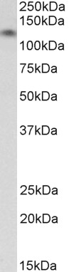
. Electrophoresis was performed on a 5-20% SDS-PAGE gel at 70V (Stacking gel) / 90V (Resolving gel) for 2-3 hours. The sample well of each lane was loaded with 50ug of sample under reducing conditions. Lane 1: human U87 whole cell lysates, Lane 2: human HELA whole cell lysates. After Electrophoresis, proteins were transferred to a Nitrocellulose membrane at 150mA for 50-90 minutes. Blocked the membrane with 5% Non-fat Milk/ TBS for 1.5 hour at RT. The membrane was incubated with rabbit anti-H Cadherin/CDH13 antigen affinity purified polyclonal antibody (Catalog # A01986-1) at 0.5 microg/mL overnight at 4°C, then washed with TBS-0.1%Tween 3 times with 5 minutes each and probed with a goat anti-rabbit IgG-HRP secondary antibody at a dilution of 1:5000 for 1.5 hour at RT. The signal is developed using an Enhanced Chemiluminescent detection (ECL) kit (Catalog # EK1002) with Tanon 5200 system. A specific band was detected for H Cadherin/CDH13 at approximately 105KD,130KD. The expected band size for H Cadherin/CDH13 is at 78KD.")
. H Cadherin/CDH13 was detected in paraffin-embedded section of human appendicitis tissue. Heat mediated antigen retrieval was performed in EDTA buffer (pH8.0, epitope retrieval solution). The tissue section was blocked with 10% goat serum. The tissue section was then incubated with 2microg/ml rabbit anti-H Cadherin/CDH13 Antibody (A01986-1) overnight at 4°C. Biotinylated goat anti-rabbit IgG was used as secondary antibody and incubated for 30 minutes at 37°C. The tissue section was developed using Strepavidin-Biotin-Complex (SABC) (Catalog # SA1022) with DAB as the chromogen.")
. H Cadherin/CDH13 was detected in paraffin-embedded section of human gastric cancer tissue. Heat mediated antigen retrieval was performed in EDTA buffer (pH8.0, epitope retrieval solution). The tissue section was blocked with 10% goat serum. The tissue section was then incubated with 2microg/ml rabbit anti-H Cadherin/CDH13 Antibody (A01986-1) overnight at 4°C. Biotinylated goat anti-rabbit IgG was used as secondary antibody and incubated for 30 minutes at 37°C. The tissue section was developed using Strepavidin-Biotin-Complex (SABC) (Catalog # SA1022) with DAB as the chromogen.")
. H Cadherin/CDH13 was detected in paraffin-embedded section of human renal carcinoma tissue. Heat mediated antigen retrieval was performed in EDTA buffer (pH8.0, epitope retrieval solution). The tissue section was blocked with 10% goat serum. The tissue section was then incubated with 2microg/ml rabbit anti-H Cadherin/CDH13 Antibody (A01986-1) overnight at 4°C. Biotinylated goat anti-rabbit IgG was used as secondary antibody and incubated for 30 minutes at 37°C. The tissue section was developed using Strepavidin-Biotin-Complex (SABC) (Catalog # SA1022) with DAB as the chromogen.")
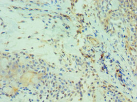
. H Cadherin/CDH13 was detected in paraffin-embedded section of human melanoma tissue. Heat mediated antigen retrieval was performed in EDTA buffer (pH8.0, epitope retrieval solution). The tissue section was blocked with 10% goat serum. The tissue section was then incubated with 2microg/ml rabbit anti-H Cadherin/CDH13 Antibody (A01986-1) overnight at 4°C. Biotinylated goat anti-rabbit IgG was used as secondary antibody and incubated for 30 minutes at 37°C. The tissue section was developed using Strepavidin-Biotin-Complex (SABC) (Catalog # SA1022) with DAB as the chromogen.")
. H Cadherin/CDH13 was detected in paraffin-embedded section of human ovarian cancer tissue. Heat mediated antigen retrieval was performed in EDTA buffer (pH8.0, epitope retrieval solution). The tissue section was blocked with 10% goat serum. The tissue section was then incubated with 2microg/ml rabbit anti-H Cadherin/CDH13 Antibody (A01986-1) overnight at 4°C. Biotinylated goat anti-rabbit IgG was used as secondary antibody and incubated for 30 minutes at 37°C. The tissue section was developed using Strepavidin-Biotin-Complex (SABC) (Catalog # SA1022) with DAB as the chromogen.")
. H Cadherin/CDH13 was detected in paraffin-embedded section of human skeletal muscles tissue. Heat mediated antigen retrieval was performed in EDTA buffer (pH8.0, epitope retrieval solution). The tissue section was blocked with 10% goat serum. The tissue section was then incubated with 2microg/ml rabbit anti-H Cadherin/CDH13 Antibody (A01986-1) overnight at 4°C. Biotinylated goat anti-rabbit IgG was used as secondary antibody and incubated for 30 minutes at 37°C. The tissue section was developed using Strepavidin-Biotin-Complex (SABC) (Catalog # SA1022) with DAB as the chromogen.")
. H Cadherin/CDH13 was detected in paraffin-embedded section of human skin cancer tissue. Heat mediated antigen retrieval was performed in EDTA buffer (pH8.0, epitope retrieval solution). The tissue section was blocked with 10% goat serum. The tissue section was then incubated with 2microg/ml rabbit anti-H Cadherin/CDH13 Antibody (A01986-1) overnight at 4°C. Biotinylated goat anti-rabbit IgG was used as secondary antibody and incubated for 30 minutes at 37°C. The tissue section was developed using Strepavidin-Biotin-Complex (SABC) (Catalog # SA1022) with DAB as the chromogen.")
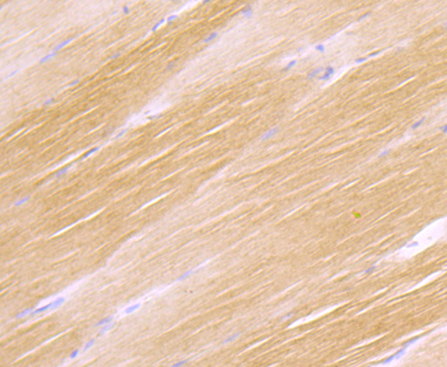
. H Cadherin/CDH13 was detected in paraffin-embedded section of rat cardiac muscle tissue. Heat mediated antigen retrieval was performed in EDTA buffer (pH8.0, epitope retrieval solution). The tissue section was blocked with 10% goat serum. The tissue section was then incubated with 2microg/ml rabbit anti-H Cadherin/CDH13 Antibody (A01986-1) overnight at 4°C. Biotinylated goat anti-rabbit IgG was used as secondary antibody and incubated for 30 minutes at 37°C. The tissue section was developed using Strepavidin-Biotin-Complex (SABC) (Catalog # SA1022) with DAB as the chromogen.")
Figure 10. Flow Cytometry analysis of HELA cells using anti-H Cadherin/CDH13 antibody (A01986-1). Overlay histogram showing HELA cells stained with A01986-1 (Blue line). The cells were fixed with 4% paraformaldehyde and blocked with 10% normal goat serum. And then incubated with rabbit anti-H Cadherin/CDH13 Antibody (A01986-1, 1microg/1x106 cells) for 30 min at 20°C. DyLight®488 conjugated goat anti-rabbit IgG (BA1127, 5-10microg/1x106 cells) was used as secondary antibody for 30 minutes at 20°C. Isotype control antibody (Green line) was rabbit IgG (1microg/1x106) used under the same conditions. Unlabelled sample without incubation with primary antibody and secondary antibody (Red line) was used as a blank control.
Anti-H Cadherin/CDH13 Antibody Picoband(r)
A01986-1-CARRIER-FREE
ApplicationsFlow Cytometry, ImmunoFluorescence, Western Blot, ELISA, ImmunoCytoChemistry, ImmunoHistoChemistry
Product group Antibodies
ReactivityHuman, Rat
TargetCDH13
Overview
- SupplierBoster Bio
- Product NameAnti-H Cadherin/CDH13 Antibody Picoband(r)
- Delivery Days Customer9
- ApplicationsFlow Cytometry, ImmunoFluorescence, Western Blot, ELISA, ImmunoCytoChemistry, ImmunoHistoChemistry
- CertificationResearch Use Only
- ClonalityPolyclonal
- Concentration500 ug/ml
- Gene ID1012
- Target nameCDH13
- Target descriptioncadherin 13
- Target synonymsCDHH, P105, cadherin-13, H-cadherin (heart), T-cad, T-cadherin, cadherin 13, H-cadherin (heart), heart cadherin
- HostRabbit
- IsotypeIgG
- Protein IDP55290
- Protein NameCadherin-13
- Scientific DescriptionBoster Bio Anti-H Cadherin/CDH13 Antibody Picoband® catalog # A01986-1. Tested in ELISA, Flow Cytometry, IF, IHC, ICC, WB applications. This antibody reacts with Human, Rat. The brand Picoband indicates this is a premium antibody that guarantees superior quality, high affinity, and strong signals with minimal background in Western blot applications. Only our best-performing antibodies are designated as Picoband, ensuring unmatched performance.
- ReactivityHuman, Rat
- Storage Instruction-20°C,2°C to 8°C
- UNSPSC12352203
Related products
Product group Antibodies
Anti-H Cadherin AntibodyA82654
ApplicationsWestern Blot, ELISA
ReactivityHuman
- SizePrice
Product group Antibodies

H Cadherin AntibodyABX027987
ApplicationsFlow Cytometry, Western Blot, ELISA, ImmunoHistoChemistry
- SizePrice
Product group Antibodies
Anti-CDH13 AntibodyAMAB92034
ApplicationsWestern Blot, ImmunoHistoChemistry
ReactivityHuman
TargetCDH13
- SizePrice
Product group Antibodies
H Cadherin Recombinant AntibodyBSM-54321R
ApplicationsImmunoFluorescence, ImmunoHistoChemistry, ImmunoHistoChemistry Frozen, ImmunoHistoChemistry Paraffin
ReactivityHuman, Mouse, Rat
TargetCDH13
- SizePrice
Product group Antibodies

Goat anti-CDH13 / H-cadherinEB12228
ApplicationsWestern Blot, ELISA, ImmunoHistoChemistry
ReactivityHuman
TargetCDH13
- SizePrice
Product group Antibodies
ApplicationsImmunoPrecipitation, Western Blot, ImmunoCytoChemistry, ImmunoHistoChemistry
ReactivityMouse, Porcine, Rat
TargetCDH13
- SizePrice
Product group Antibodies
CDH13 AntibodyCSB-PA005039ESR1HU
ApplicationsELISA, ImmunoHistoChemistry
ReactivityHuman
TargetCDH13
- SizePrice
![IHC-P analysis of human esophageal tissue section using GTX02852 CDH13 antibody [GT1255]. Dilution : 1:100](https://www.genetex.com/upload/website/prouct_img/normal/GTX02852/A3244_IHC_02_(1073419)_w_23053123_883.webp)
Product group Antibodies
CDH13 antibody [GT1255]GTX02852
ApplicationsWestern Blot, ImmunoHistoChemistry, ImmunoHistoChemistry Paraffin
ReactivityHuman, Mouse, Rat
TargetCDH13
- SizePrice
Product group Antibodies

CDH13 / Cadherin 13 AntibodyLS-C331675
ApplicationsWestern Blot, ImmunoHistoChemistry
ReactivityHuman, Mouse
TargetCDH13
- SizePrice