

Whole cell extract (30 μg) was separated by 12% SDS-PAGE, and the membrane was blotted with CDK4 antibody (GTX102993) diluted at 1:500. The HRP-conjugated anti-rabbit IgG antibody (GTX213110-01) was used to detect the primary antibody.
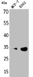
 were separated by 12% SDS-PAGE, and the membrane was blotted with CDK4 antibody (GTX102993) diluted at 1:1000. The HRP-conjugated anti-rabbit IgG antibody (GTX213110-01) was used to detect the primary antibody.")
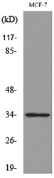
 was separated by 12% SDS-PAGE, and the membrane was blotted with CDK4 antibody (GTX102993) diluted at 1:500. The HRP-conjugated anti-rabbit IgG antibody (GTX213110-01) was used to detect the primary antibody.")
 and transfected (+) 293T whole cell extracts (30 μg) were separated by 12% SDS-PAGE, and the membrane was blotted with CDK4 antibody (GTX102993) diluted at 1:1000. The HRP-conjugated anti-rabbit IgG antibody (GTX213110-01) was used to detect the primary antibody.")
 antibody at 1:500 dilution.
Antigen Retrieval: Trilogy? (EDTA based, pH 8.0) buffer, 15min")
![CDK4 antibody detects CDK4 protein at nucleus by immunofluorescent analysis. Sample: MCF7 cells were fixed in 4% paraformaldehyde at RT for 15 min. Green: CDK4 protein stained by CDK4 antibody (GTX102993) diluted at 1:1000. Red: p21 Cip1, a nucleus marker, stained by p21 Cip1 antibody [GT1032] (GTX629543) diluted at 1:500. Blue: Hoechst 33342 staining.](https://www.genetex.com/upload/website/prouct_img/normal/GTX102993/GTX102993_39883_20150518_IFA_w_23060119_634.webp "CDK4 antibody detects CDK4 protein at nucleus by immunofluorescent analysis. Sample: MCF7 cells were fixed in 4% paraformaldehyde at RT for 15 min. Green: CDK4 protein stained by CDK4 antibody (GTX102993) diluted at 1:1000. Red: p21 Cip1, a nucleus marker, stained by p21 Cip1 antibody [GT1032] (GTX629543) diluted at 1:500. Blue: Hoechst 33342 staining.")
 was separated by 12% SDS-PAGE, and the membrane was blotted with CDK4 antibody (GTX102993) diluted at 1:500. The HRP-conjugated anti-rabbit IgG antibody (GTX213110-01) was used to detect the primary antibody.")
 diluted at 1:500. Red: phalloidin, a cytoskeleton marker, diluted at 1:200. Scale bar = 10 μm.")


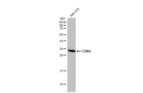
Whole cell extract (30 μg) was separated by 12% SDS-PAGE, and the membrane was blotted with CDK4 antibody (GTX102993) diluted at 1:500. The HRP-conjugated anti-rabbit IgG antibody (GTX213110-01) was used to detect the primary antibody.
CDK4 antibody
GTX102993
ApplicationsImmunoFluorescence, Western Blot, ImmunoCytoChemistry, ImmunoHistoChemistry, ImmunoHistoChemistry Paraffin
Product group Antibodies
ReactivityBovine, Human, Mouse, Rat
TargetCDK4
Overview
- SupplierGeneTex
- Product NameCDK4 antibody
- Delivery Days Customer9
- Application Supplier NoteWB: 1:500-1:3000. ICC/IF: 1:100-1:1000. IHC-P: 1:100-1:1000. *Optimal dilutions/concentrations should be determined by the researcher.Not tested in other applications.
- ApplicationsImmunoFluorescence, Western Blot, ImmunoCytoChemistry, ImmunoHistoChemistry, ImmunoHistoChemistry Paraffin
- CertificationResearch Use Only
- ClonalityPolyclonal
- Concentration0.82 mg/ml
- ConjugateUnconjugated
- Gene ID1019
- Target nameCDK4
- Target descriptioncyclin dependent kinase 4
- Target synonymsCMM3, PSK-J3, cyclin-dependent kinase 4, cell division protein kinase 4
- HostRabbit
- IsotypeIgG
- Protein IDP11802
- Protein NameCyclin-dependent kinase 4
- Scientific DescriptionThe protein encoded by this gene is a member of the Ser/Thr protein kinase family. This protein is highly similar to the gene products of S. cerevisiae cdc28 and S. pombe cdc2. It is a catalytic subunit of the protein kinase complex that is important for cell cycle G1 phase progression. The activity of this kinase is restricted to the G1-S phase, which is controlled by the regulatory subunits D-type cyclins and CDK inhibitor p16(INK4a). This kinase was shown to be responsible for the phosphorylation of retinoblastoma gene product (Rb). Mutations in this gene as well as in its related proteins including D-type cyclins, p16(INK4a) and Rb were all found to be associated with tumorigenesis of a variety of cancers. Multiple polyadenylation sites of this gene have been reported. [provided by RefSeq]
- ReactivityBovine, Human, Mouse, Rat
- Storage Instruction-20°C or -80°C,2°C to 8°C
- UNSPSC41116161
Datasheet
Related products
Product group Antibodies
Anti-CDK4 Antibody144-00366
ApplicationsImmunoFluorescence, Western Blot, ImmunoHistoChemistry
ReactivityHuman, Mouse, Rat
TargetCDK4
- SizePrice
Product group Antibodies
Anti-CDK4 AntibodyA98225
ApplicationsWestern Blot, ELISA
ReactivityHuman, Mouse, Rat
- SizePrice

Product group Antibodies
Anti-CDK4 AntibodyAMAB91499
ApplicationsWestern Blot, ImmunoCytoChemistry, ImmunoHistoChemistry
ReactivityHuman
TargetCDK4
- SizePrice
Product group Antibodies
ApplicationsWestern Blot, ImmunoHistoChemistry
ReactivityRat
TargetCDK4
- SizePrice
Product group Antibodies
CDK4 AntibodyCSB-PA005118
ApplicationsWestern Blot, ELISA
ReactivityHuman, Mouse, Rat
TargetCDK4
- SizePrice
Product group Antibodies
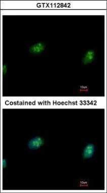
CDK4 antibodyGTX112842
ApplicationsImmunoFluorescence, Western Blot, ImmunoCytoChemistry
ReactivityHuman
TargetCDK4
- SizePrice
Product group Antibodies
CDK4 antibodyGTX16516
ApplicationsImmunoPrecipitation, Western Blot
ReactivityHuman
TargetCDK4
- SizePrice
![WB analysis of HeLa and human uterine mesodermal tumor cell-SKUT lysates using GTX26315 CDK4 antibody [DCS-31].
Dilution : 1:2000](https://www.genetex.com/upload/website/prouct_img/normal/GTX26315/GTX26315_20191017_WB_1_w_23060722_457.webp)
Product group Antibodies
CDK4 antibody [DCS-31]GTX26315
ApplicationsImmunoFluorescence, ImmunoPrecipitation, Western Blot, ImmunoCytoChemistry, ImmunoHistoChemistry, ImmunoHistoChemistry Paraffin
ReactivityHuman, Mouse, Rat
TargetCDK4
- SizePrice
Product group Antibodies
CDK4 antibodyGTX26551
ApplicationsImmunoPrecipitation, Western Blot, ELISA
ReactivityHuman, Mouse, Rat
TargetCDK4
- SizePrice