

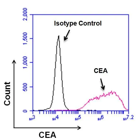
FACS analysis of BxPC-3 cells using GTX15677 CEA antibody [1106]. Pink histogram : Primary antibody Black histogram : Isotype control antibody Dilution : 10 microg/ml in PBS + 5% FCS
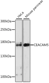
![WB analysis of 20 ug of BxPC-3, HeLa, and HepG2 whole cell lysates per well using GTX15677 CEA antibody [1106]. Dilution : 1 microg/ml](https://www.genetex.com/upload/website/prouct_img/normal/GTX15677/GTX15677_1516_WB_w_23060620_359.webp "WB analysis of 20 ug of BxPC-3, HeLa, and HepG2 whole cell lysates per well using GTX15677 CEA antibody [1106]. Dilution : 1 microg/ml")
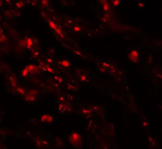
![ICC/IF analysis of BxPC-3 cells using GTX15677 CEA antibody [1106]. Green : Primary antibody Blue : Nuclei Fixation : 4% paraformaldehyde Permeabilization : 0.1% Trito X-100 for 10 minutes Dilution : 10 microg/ml in blocking buffer for 1 hour at room temperature](https://www.genetex.com/upload/website/prouct_img/normal/GTX15677/GTX15677_287_ICC-IF_w_23060620_995.webp "ICC/IF analysis of BxPC-3 cells using GTX15677 CEA antibody [1106]. Green : Primary antibody Blue : Nuclei Fixation : 4% paraformaldehyde Permeabilization : 0.1% Trito X-100 for 10 minutes Dilution : 10 microg/ml in blocking buffer for 1 hour at room temperature")
FACS analysis of BxPC-3 cells using GTX15677 CEA antibody [1106]. Pink histogram : Primary antibody Black histogram : Isotype control antibody Dilution : 10 microg/ml in PBS + 5% FCS
CD66e antibody [1106]
GTX15677
ApplicationsFlow Cytometry, ImmunoFluorescence, ImmunoPrecipitation, Western Blot, ELISA, ImmunoCytoChemistry, RadioImmunoAssay
Product group Antibodies
TargetCEACAM5
Overview
- SupplierGeneTex
- Product NameCD66e antibody [1106]
- Delivery Days Customer9
- Application Supplier NoteWB: 1microg/ml. FACS: 5-20microg/ml. *Optimal dilutions/concentrations should be determined by the researcher.Not tested in other applications.
- ApplicationsFlow Cytometry, ImmunoFluorescence, ImmunoPrecipitation, Western Blot, ELISA, ImmunoCytoChemistry, RadioImmunoAssay
- CertificationResearch Use Only
- ClonalityMonoclonal
- Clone ID1106
- Concentration1 mg/ml
- ConjugateUnconjugated
- Gene ID1048
- Target nameCEACAM5
- Target descriptionCEA cell adhesion molecule 5
- Target synonymsCD66e, CEA, cell adhesion molecule CEACAM5, carcinoembryonic antigen related cell adhesion molecule 5, meconium antigen 100
- HostMouse
- IsotypeIgG1
- Protein IDP06731
- Protein NameCell adhesion molecule CEACAM5
- Scientific DescriptionThis gene encodes a cell surface glycoprotein that represents the founding member of the carcinoembryonic antigen (CEA) family of proteins. The encoded protein is used as a clinical biomarker for gastrointestinal cancers and may promote tumor development through its role as a cell adhesion molecule. Additionally, the encoded protein may regulate differentiation, apoptosis, and cell polarity. This gene is present in a CEA family gene cluster on chromosome 19. Alternative splicing results in multiple transcript variants. [provided by RefSeq, Jul 2015]
- Storage Instruction-20°C or -80°C,2°C to 8°C
- UNSPSC12352203
Datasheet
Related products
Product group Antibodies
CD66e antibodyGTX31551
ApplicationsWestern Blot, ELISA, ImmunoHistoChemistry, ImmunoHistoChemistry Paraffin
TargetCEACAM5
- SizePrice
Product group Antibodies
CD66e antibody [N1N3]GTX113161
ApplicationsWestern Blot, ImmunoHistoChemistry, ImmunoHistoChemistry Paraffin
TargetCEACAM5
- SizePrice
![IHC-P analysis of human bowel tissue using GTX01863 CD66e antibody [12-140-10]. Note cytoplasmic staining of epithelial cells.](https://www.genetex.com/upload/website/prouct_img/normal/GTX01863/GTX01863_20200811_IHC-P_24_w_23053121_712.webp)
Product group Antibodies
CD66e antibody [12-140-10]GTX01863
ApplicationsImmunoHistoChemistry, ImmunoHistoChemistry Frozen, ImmunoHistoChemistry Paraffin
TargetCEACAM5
- SizePrice
Product group Antibodies
CD66e antibody [HL2467]GTX638816
ApplicationsWestern Blot, ImmunoHistoChemistry, ImmunoHistoChemistry Paraffin
TargetCEACAM5
- SizePrice
Product group Antibodies
CD66e antibody [C365D3(NCRC23)]GTX75309
ApplicationsFlow Cytometry, ELISA, ImmunoHistoChemistry, ImmunoHistoChemistry Frozen, ImmunoHistoChemistry Paraffin
TargetCEACAM5
- SizePrice
Product group Antibodies
ApplicationsFlow Cytometry
TargetCEACAM5
- SizePrice
![IHC-P analysis of colorectal carcinoma tissue using GTX80201 CD66e antibody [CB30]. Diultion : 10microg/ml](https://www.genetex.com/upload/website/prouct_img/normal/GTX80201/GTX80201_20191025_AP_003_422_w_23061322_861.webp)
Product group Antibodies
CD66e antibody [CB30]GTX80201
ApplicationsFlow Cytometry, ImmunoPrecipitation, ImmunoHistoChemistry, ImmunoHistoChemistry Frozen, ImmunoHistoChemistry Paraffin
TargetCEACAM5
- SizePrice
Product group Antibodies
CD66e antibody [CB30]GTX41869
ApplicationsFlow Cytometry, ImmunoPrecipitation, ImmunoHistoChemistry, ImmunoHistoChemistry Frozen, ImmunoHistoChemistry Paraffin
TargetCEACAM5
- SizePrice