

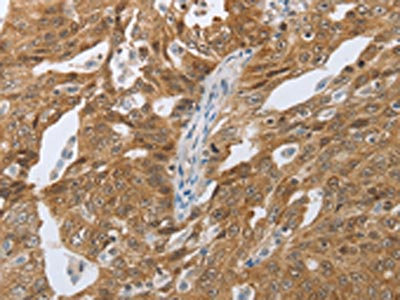
The image on the left is immunohistochemistry of paraffin-embedded Human tonsil tissue using CSB-PA981539(CEBPA Antibody) at dilution 1/40, on the right is treated with synthetic peptide. (Original magnification: x200)
 at dilution 1/40, on the right is treated with synthetic peptide. (Original magnification: x200)")
The image on the left is immunohistochemistry of paraffin-embedded Human tonsil tissue using CSB-PA981539(CEBPA Antibody) at dilution 1/40, on the right is treated with synthetic peptide. (Original magnification: x200)
CEBPA Antibody
CSB-PA981539
ApplicationsELISA, ImmunoHistoChemistry
Product group Antibodies
TargetCEBPA
Overview
- SupplierCusabio
- Product NameCEBPA Antibody
- Delivery Days Customer20
- ApplicationsELISA, ImmunoHistoChemistry
- CertificationResearch Use Only
- ClonalityPolyclonal
- ConjugateUnconjugated
- Gene ID1050
- Target nameCEBPA
- Target descriptionCCAAT enhancer binding protein alpha
- Target synonymsC/EBP-alpha; CCAAT/enhancer binding protein (C/EBP), alpha; CCAAT/enhancer-binding protein alpha; CEBP
- HostRabbit
- IsotypeIgG
- Protein IDP49715
- Protein NameCCAAT/enhancer-binding protein alpha
- Scientific DescriptionThe protein encoded by this intronless gene is a bZIP transcription factor which can bind as a homodimer to certain promoters and enhancers. It can also form heterodimers with the related proteins CEBP-beta and CEBP-gamma. The encoded protein has been shown to bind to the promoter and modulate the expression of the gene encoding leptin, a protein that plays an important role in body weight homeostasis. Also, the encoded protein can interact with CDK2 and CDK4, thereby inhibiting these kinases and causing growth arrest in cultured cells.
- Storage Instruction-20°C or -80°C
- UNSPSC12352203
Related products
Product group Antibodies
CEBPA Polyclonal AntibodyCAC14713
ApplicationsWestern Blot, ELISA, ImmunoHistoChemistry
TargetCEBPA
- SizePrice
Product group Antibodies
References
C/EBP alpha antibody [N1], N-termGTX100674
ApplicationsImmunoPrecipitation, Western Blot, ChIP Chromatin ImmunoPrecipitation, ImmunoHistoChemistry, ImmunoHistoChemistry Paraffin
TargetCEBPA
- SizePrice
Product group Antibodies
Anti-CEBPA AntibodyHPA052734
ApplicationsImmunoCytoChemistry
TargetCEBPA
- SizePrice
Product group Antibodies
CEBPA AntibodyCSB-PA001073
ApplicationsWestern Blot, ELISA, ImmunoHistoChemistry
TargetCEBPA
- SizePrice
Product group Antibodies
Anti-CEBPA Antibody144-61658
ApplicationsWestern Blot, ImmunoHistoChemistry
TargetCEBPA
- SizePrice
Product group Antibodies

C/EBP Alpha / CEBPA AntibodyLS-C761164
ApplicationsWestern Blot, ImmunoHistoChemistry
TargetCEBPA
- SizePrice
Product group Antibodies
References
CEBP-alpha Polyclonal AntibodyBS-1630R
ApplicationsFlow Cytometry, ImmunoFluorescence, Western Blot, ELISA, ImmunoCytoChemistry, ImmunoHistoChemistry, ImmunoHistoChemistry Frozen, ImmunoHistoChemistry Paraffin
TargetCEBPA
- SizePrice

Product group Antibodies
Anti-CEBP Alpha/CEBPA Antibody Picoband(r)A00386-1-CARRIER-FREE
ApplicationsFlow Cytometry, ImmunoFluorescence, Western Blot, ELISA, ImmunoCytoChemistry
TargetCEBPA
- SizePrice