

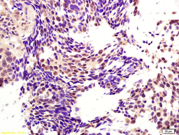
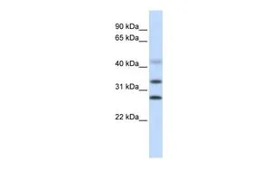
CENPH / CENP-H Antibody (clone OTI4G6)
LS-C173490
ApplicationsFlow Cytometry, Western Blot
Product group Antibodies
TargetCENPH
Overview
- SupplierLifeSpan BioSciences
- Product NameCENPH / CENP-H Antibody (clone OTI4G6)
- Delivery Days Customer23
- ApplicationsFlow Cytometry, Western Blot
- Applications SupplierFlo (1:100), WB (1:500)
- CertificationResearch Use Only
- ClonalityMonoclonal
- Clone IDOTI4G6
- Concentration0.42 mg/ml
- ConjugateUnconjugated
- Estimated Purity...
- Gene ID64946
- Target nameCENPH
- Target descriptioncentromere protein H
- Target synonymsCENP-H; centromere protein H; interphase centromere complex protein 35; kinetochore protein CENP-H
- HostMouse
- IsotypeIgG1
- Storage Instruction-20°C
- UNSPSC12352203
Related products
Product group Antibodies
CENPH Polyclonal AntibodyBS-6519R
ApplicationsImmunoFluorescence, Western Blot, ELISA, ImmunoCytoChemistry, ImmunoHistoChemistry, ImmunoHistoChemistry Frozen, ImmunoHistoChemistry Paraffin
TargetCENPH
- SizePrice
Product group Antibodies
ApplicationsWestern Blot, ImmunoHistoChemistry
TargetCENPH
- SizePrice

Product group Antibodies
Anti-CENPH Antibody Picoband(r)A06302-3-CARRIER-FREE
ApplicationsFlow Cytometry, Western Blot, ELISA
TargetCENPH
- SizePrice

Product group Antibodies
Anti-CENPH (Center) Antibody102-27680
ApplicationsFlow Cytometry, Western Blot, ImmunoHistoChemistry, ImmunoHistoChemistry Paraffin
TargetCENPH
- SizePrice
Product group Antibodies
CENPH antibody, InternalGTX44860
ApplicationsWestern Blot
TargetCENPH
- SizePrice
Product group Antibodies

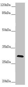
CENPH / CENP-H AntibodyLS-C409908
ApplicationsWestern Blot
TargetCENPH
- SizePrice

Product group Antibodies
Anti-CENPH AntibodyHPA036494
ApplicationsWestern Blot, ImmunoCytoChemistry, ImmunoHistoChemistry
TargetCENPH
- SizePrice
Product group Antibodies
CENPH AntibodyCSB-PA005211ESR1HU
ApplicationsWestern Blot, ELISA, ImmunoHistoChemistry
ReactivityHuman, Mouse
TargetCENPH
- SizePrice