

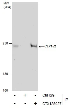
Immunoprecipitation of CEP152 protein from HeLa whole cell extracts using 5 μg of CEP152 antibody (GTX128027). Western blot analysis was performed using CEP152 antibody (GTX128027). EasyBlot anti-Rabbit IgG (GTX221666-01) was used as a secondary reagent.
 diluted at 1:2000.
Red: Tubulin, alpha 1b protein stained by TUBA1B antibody (GTX11304) diluted at 1:5000.
Blue: Hoechst 33342 staining.")
 were separated by 5% SDS-PAGE, and the membrane was blotted with CEP152 antibody (GTX128027) diluted at 1:500. The HRP-conjugated anti-rabbit IgG antibody (GTX213110-01) was used to detect the primary antibody. Corresponding RNA expression data for the same cell lines are based on Human Protein Atlas program.")
![CEP152 antibody detects CEP152 protein at cytoskeleton and cytoplasm by immunofluorescent analysis. Sample: HeLa cells were fixed in MeOH. Green: CEP152 stained by CEP152 antibody (GTX128027) diluted at 1:500. Red: alpha Tubulin, a cytoskeleton marker, stained by alpha Tubulin antibody [GT114] (GTX628802) diluted at 1:1000. Blue: Fluoroshield with DAPI (GTX30920).](https://www.genetex.com/upload/website/prouct_img/normal/GTX128027/GTX128027_45126_20240105_ICC_IF_24011618_149.webp "CEP152 antibody detects CEP152 protein at cytoskeleton and cytoplasm by immunofluorescent analysis. Sample: HeLa cells were fixed in MeOH. Green: CEP152 stained by CEP152 antibody (GTX128027) diluted at 1:500. Red: alpha Tubulin, a cytoskeleton marker, stained by alpha Tubulin antibody [GT114] (GTX628802) diluted at 1:1000. Blue: Fluoroshield with DAPI (GTX30920).")
Immunoprecipitation of CEP152 protein from HeLa whole cell extracts using 5 μg of CEP152 antibody (GTX128027). Western blot analysis was performed using CEP152 antibody (GTX128027). EasyBlot anti-Rabbit IgG (GTX221666-01) was used as a secondary reagent.
CEP152 antibody
GTX128027
ApplicationsImmunoFluorescence, ImmunoPrecipitation, Western Blot, ImmunoCytoChemistry, ImmunoHistoChemistry, ImmunoHistoChemistry Frozen
Product group Antibodies
ReactivityHuman, Mouse
TargetCEP152
Overview
- SupplierGeneTex
- Product NameCEP152 antibody
- Delivery Days Customer9
- Application Supplier NoteWB: 1:500-1:3000. ICC/IF: 1:100-1:1000. IP: 1:100-1:500. *Optimal dilutions/concentrations should be determined by the researcher.Not tested in other applications.
- ApplicationsImmunoFluorescence, ImmunoPrecipitation, Western Blot, ImmunoCytoChemistry, ImmunoHistoChemistry, ImmunoHistoChemistry Frozen
- CertificationResearch Use Only
- ClonalityPolyclonal
- Concentration1.34 mg/ml
- ConjugateUnconjugated
- Gene ID22995
- Target nameCEP152
- Target descriptioncentrosomal protein 152
- Target synonymsMCPH4, MCPH9, SCKL5, centrosomal protein of 152 kDa, asterless, centrosomal protein 152kDa, microcephaly, primary autosomal recessive 4
- HostRabbit
- IsotypeIgG
- Protein IDO94986
- Protein NameCentrosomal protein of 152 kDa
- Scientific DescriptionThis gene encodes a protein that is thought to be involved with centrosome function. Mutations in this gene have been associated with primary microcephaly (MCPH4). Alternative splicing results in multiple transcript variants. [provided by RefSeq]
- ReactivityHuman, Mouse
- Storage Instruction-20°C or -80°C,2°C to 8°C
- UNSPSC12352203
References
- Aziz K, Sieben CJ, Jeganathan KB, et al. Mosaic-variegated aneuploidy syndrome mutation or haploinsufficiency in Cep57 impairs tumor suppression. J Clin Invest. 2018,128(8):3517-3534. doi: 10.1172/JCI120316Read this paper
Datasheet
Related products

Product group Antibodies
Anti-CEP152 AntibodyA30605
ApplicationsImmunoFluorescence, Western Blot, ELISA, ImmunoCytoChemistry, ImmunoHistoChemistry
TargetCEP152
- SizePrice
![CEP152 antibody [GT1315] detects CEP152 protein by western blot analysis. Various whole cell extracts (30 μg) were separated by 5 % SDS-PAGE, and blotted with CEP152 antibody [GT1315] (GTX630984) diluted by 1:500, and developed with Trident femto Western HRP Substrate (GTX14698).](https://www.genetex.com/upload/website/prouct_img/normal/GTX630984/GTX630984_41638_WB_w_23061202_171.webp)
Product group Antibodies
CEP152 antibody [GT1315]GTX630984
ApplicationsImmunoFluorescence, Western Blot, ImmunoCytoChemistry
ReactivityHuman
TargetCEP152
- SizePrice
![CEP152 antibody [GT1285] detects CEP152 protein by western blot analysis. Whole cell extracts (30 μg) was separated by 5 % SDS-PAGE, and blotted with CEP152 antibody [GT1285] (GTX631486) diluted by 1:500, and developed with Trident femto Western HRP Substrate (GTX14698).](https://www.genetex.com/upload/website/prouct_img/normal/GTX631486/GTX631486_41750_WB_Trident-Sharp-ECL_w_23061202_429.webp)
Product group Antibodies
CEP152 antibody [GT1285]GTX631486
ApplicationsWestern Blot
ReactivityHuman
TargetCEP152
- SizePrice
Product group Antibodies
CEP152 Polyclonal AntibodyBS-7787R
ApplicationsImmunoFluorescence, Western Blot, ELISA, ImmunoCytoChemistry, ImmunoHistoChemistry, ImmunoHistoChemistry Frozen, ImmunoHistoChemistry Paraffin
ReactivityCanine, Equine, Human, Mouse, Rat
TargetCEP152
- SizePrice
Product group Antibodies
Anti-CEP152 AntibodyA45350
ApplicationsImmunoHistoChemistry
ReactivityHuman, Mouse
- SizePrice
![Various whole cell extracts (30 μg) were separated by 5% SDS-PAGE, and the membrane was blotted with CEP152 antibody [HL2763] (GTX639628) diluted at 1:1000. The HRP-conjugated anti-rabbit IgG antibody (GTX213110-01) was used to detect the primary antibody, and the signal was developed with Trident ECL plus-Enhanced. Corresponding RNA expression data for the same cell lines are based on Human Protein Atlas program.](https://www.genetex.com/upload/website/prouct_img/normal/GTX639628/GTX639628_45362_20240329_WB_TPM_watermark_24041019_848.webp)
Product group Antibodies
CEP152 antibody [HL2763]GTX639628
ApplicationsImmunoFluorescence, Western Blot, ImmunoCytoChemistry
ReactivityHuman
TargetCEP152
- SizePrice
![CEP152 antibody [HL2884] detects CEP152 protein at centrosome by immunofluorescent analysis. Sample: HeLa cells were fixed in 4% paraformaldehyde at RT for 15 min. Green: CEP152 stained by CEP152 antibody [HL2884] (GTX640163) diluted at 1:500. Red: alpha Tubulin, a cytoskeleton marker, stained by alpha Tubulin antibody [GT114] (GTX628802) diluted at 1:1000. Blue: Fluoroshield with DAPI (GTX30920).](https://www.genetex.com/upload/website/prouct_img/normal/GTX640163/GTX640163_T-45369_20240426_ICC_IF_24051400_694.webp)
Product group Antibodies
CEP152 antibody [HL2884]GTX640163
ApplicationsImmunoFluorescence, Western Blot, ImmunoCytoChemistry
ReactivityHuman
TargetCEP152
- SizePrice
Product group Antibodies
CEP152 antibodyGTX87714
ApplicationsImmunoHistoChemistry, ImmunoHistoChemistry Paraffin
ReactivityHuman
TargetCEP152
- SizePrice
Product group Antibodies
CEP152 AntibodyCSB-PA008298
ApplicationsWestern Blot, ELISA, ImmunoHistoChemistry
ReactivityHuman, Mouse
TargetCEP152
- SizePrice