

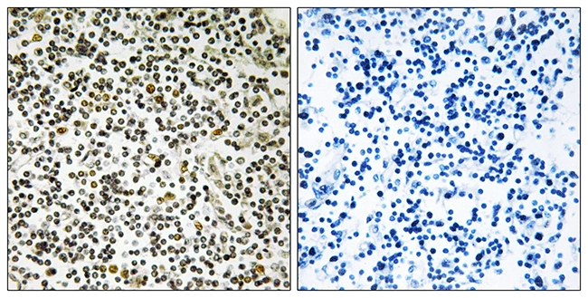
IHC-P analysis of human lymph node tissue using GTX87714 CEP152 antibody. The picture on the right is blocked with the synthesized peptide.
IHC-P analysis of human lymph node tissue using GTX87714 CEP152 antibody. The picture on the right is blocked with the synthesized peptide.
CEP152 antibody
GTX87714
ApplicationsImmunoHistoChemistry, ImmunoHistoChemistry Paraffin
Product group Antibodies
ReactivityHuman
TargetCEP152
Overview
- SupplierGeneTex
- Product NameCEP152 antibody
- Delivery Days Customer9
- Application Supplier NoteIHC-P: 1:50~1:100. *Optimal dilutions/concentrations should be determined by the researcher.Not tested in other applications.
- ApplicationsImmunoHistoChemistry, ImmunoHistoChemistry Paraffin
- CertificationResearch Use Only
- ClonalityPolyclonal
- ConjugateUnconjugated
- Gene ID22995
- Target nameCEP152
- Target descriptioncentrosomal protein 152
- Target synonymsMCPH4, MCPH9, SCKL5, centrosomal protein of 152 kDa, asterless, centrosomal protein 152kDa, microcephaly, primary autosomal recessive 4
- HostRabbit
- IsotypeIgG
- Protein IDO94986
- Protein NameCentrosomal protein of 152 kDa
- Scientific DescriptionThis gene encodes a protein that is thought to be involved with centrosome function. Mutations in this gene have been associated with primary microcephaly (MCPH4). Alternative splicing results in multiple transcript variants. [provided by RefSeq, Aug 2010]
- ReactivityHuman
- Storage Instruction-20°C or -80°C,2°C to 8°C
- UNSPSC12352203
Datasheet
Related products

Product group Antibodies
Anti-CEP152 AntibodyA30605
ApplicationsImmunoFluorescence, Western Blot, ELISA, ImmunoCytoChemistry, ImmunoHistoChemistry
TargetCEP152
- SizePrice
Product group Antibodies
References
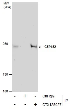
CEP152 antibodyGTX128027
ApplicationsImmunoFluorescence, ImmunoPrecipitation, Western Blot, ImmunoCytoChemistry, ImmunoHistoChemistry, ImmunoHistoChemistry Frozen
ReactivityHuman, Mouse
TargetCEP152
- SizePrice
![CEP152 antibody [GT1315] detects CEP152 protein by western blot analysis. Various whole cell extracts (30 μg) were separated by 5 % SDS-PAGE, and blotted with CEP152 antibody [GT1315] (GTX630984) diluted by 1:500, and developed with Trident femto Western HRP Substrate (GTX14698).](https://www.genetex.com/upload/website/prouct_img/normal/GTX630984/GTX630984_41638_WB_w_23061202_171.webp)
Product group Antibodies
CEP152 antibody [GT1315]GTX630984
ApplicationsImmunoFluorescence, Western Blot, ImmunoCytoChemistry
ReactivityHuman
TargetCEP152
- SizePrice
![CEP152 antibody [GT1285] detects CEP152 protein by western blot analysis. Whole cell extracts (30 μg) was separated by 5 % SDS-PAGE, and blotted with CEP152 antibody [GT1285] (GTX631486) diluted by 1:500, and developed with Trident femto Western HRP Substrate (GTX14698).](https://www.genetex.com/upload/website/prouct_img/normal/GTX631486/GTX631486_41750_WB_Trident-Sharp-ECL_w_23061202_429.webp)
Product group Antibodies
CEP152 antibody [GT1285]GTX631486
ApplicationsWestern Blot
ReactivityHuman
TargetCEP152
- SizePrice
Product group Antibodies
CEP152 Polyclonal AntibodyBS-7787R
ApplicationsImmunoFluorescence, Western Blot, ELISA, ImmunoCytoChemistry, ImmunoHistoChemistry, ImmunoHistoChemistry Frozen, ImmunoHistoChemistry Paraffin
ReactivityCanine, Equine, Human, Mouse, Rat
TargetCEP152
- SizePrice
Product group Antibodies
Anti-CEP152 AntibodyA45350
ApplicationsImmunoHistoChemistry
ReactivityHuman, Mouse
- SizePrice
![Various whole cell extracts (30 μg) were separated by 5% SDS-PAGE, and the membrane was blotted with CEP152 antibody [HL2763] (GTX639628) diluted at 1:1000. The HRP-conjugated anti-rabbit IgG antibody (GTX213110-01) was used to detect the primary antibody, and the signal was developed with Trident ECL plus-Enhanced. Corresponding RNA expression data for the same cell lines are based on Human Protein Atlas program.](https://www.genetex.com/upload/website/prouct_img/normal/GTX639628/GTX639628_45362_20240329_WB_TPM_watermark_24041019_848.webp)
Product group Antibodies
CEP152 antibody [HL2763]GTX639628
ApplicationsImmunoFluorescence, Western Blot, ImmunoCytoChemistry
ReactivityHuman
TargetCEP152
- SizePrice
![CEP152 antibody [HL2884] detects CEP152 protein at centrosome by immunofluorescent analysis. Sample: HeLa cells were fixed in 4% paraformaldehyde at RT for 15 min. Green: CEP152 stained by CEP152 antibody [HL2884] (GTX640163) diluted at 1:500. Red: alpha Tubulin, a cytoskeleton marker, stained by alpha Tubulin antibody [GT114] (GTX628802) diluted at 1:1000. Blue: Fluoroshield with DAPI (GTX30920).](https://www.genetex.com/upload/website/prouct_img/normal/GTX640163/GTX640163_T-45369_20240426_ICC_IF_24051400_694.webp)
Product group Antibodies
CEP152 antibody [HL2884]GTX640163
ApplicationsImmunoFluorescence, Western Blot, ImmunoCytoChemistry
ReactivityHuman
TargetCEP152
- SizePrice
Product group Antibodies
CEP152 AntibodyCSB-PA008298
ApplicationsWestern Blot, ELISA, ImmunoHistoChemistry
ReactivityHuman, Mouse
TargetCEP152
- SizePrice