

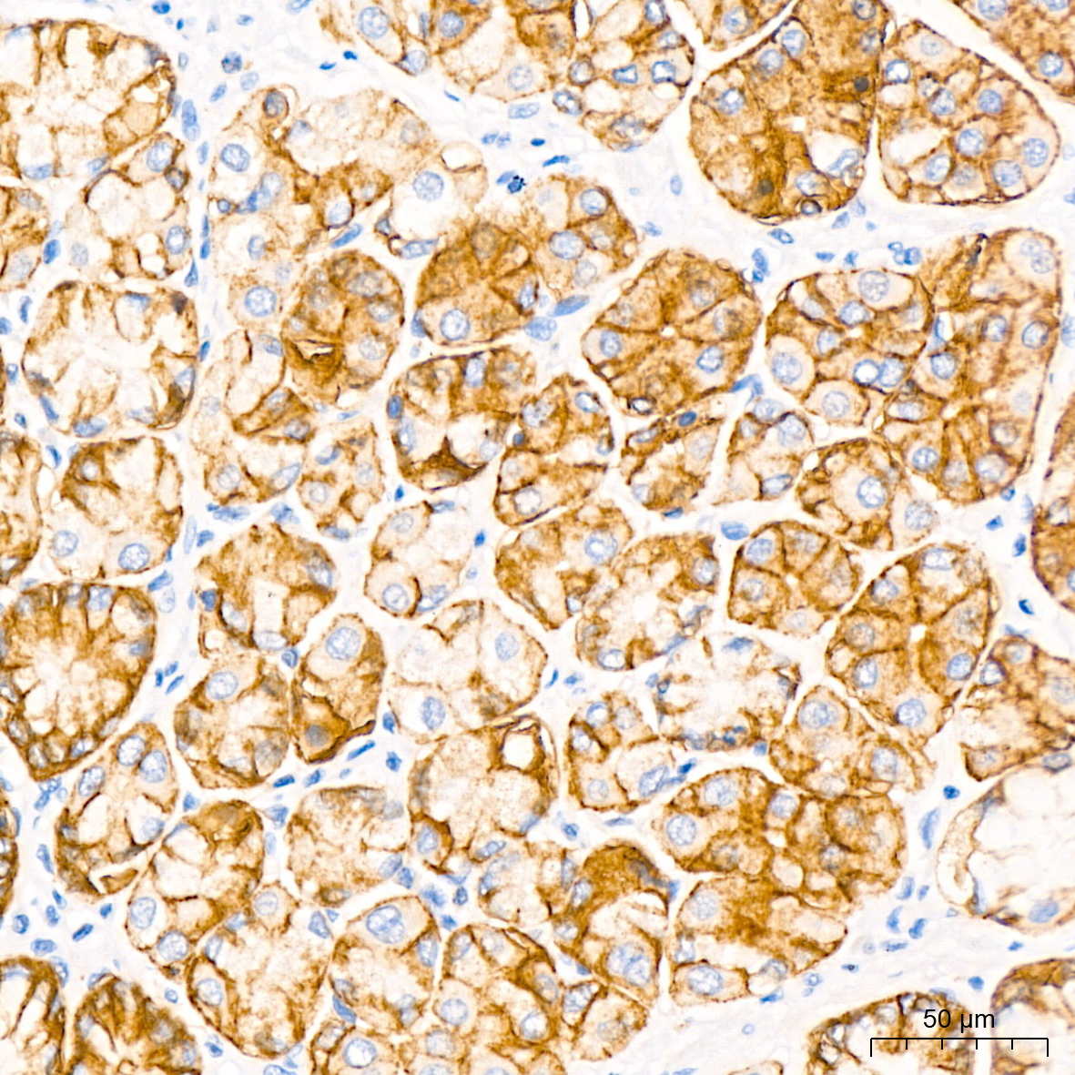
IHC-P analysis of human stomach tissue using GTX04575 Claudin 18.2 antibody [ARC5062-02]. Antigen retrieval : 10 mM Tris/EDTA buffer, pH 9.0 Dilution : 1:50
![IHC-P analysis of human stomach tissue using GTX04575 Claudin 18.2 antibody [ARC5062-02]. Antigen retrieval : 10 mM Tris/EDTA buffer, pH 9.0 Dilution : 1:50](https://www.genetex.com/upload/website/prouct_img/normal/GTX04575/GTX04575_20231024_IHC-P_23102323_471.webp "IHC-P analysis of human stomach tissue using GTX04575 Claudin 18.2 antibody [ARC5062-02]. Antigen retrieval : 10 mM Tris/EDTA buffer, pH 9.0 Dilution : 1:50")
IHC-P analysis of human stomach tissue using GTX04575 Claudin 18.2 antibody [ARC5062-02]. Antigen retrieval : 10 mM Tris/EDTA buffer, pH 9.0 Dilution : 1:50
Claudin 18.2 antibody [ARC5062-02]
GTX04575
ApplicationsImmunoHistoChemistry, ImmunoHistoChemistry Paraffin
Product group Antibodies
ReactivityHuman
TargetCLDN18
Overview
- SupplierGeneTex
- Product NameClaudin 18.2 antibody [ARC5062-02]
- Delivery Days Customer9
- Application Supplier NoteIHC-P: 1:500-1:1000. *Optimal dilutions/concentrations should be determined by the researcher.Not tested in other applications.
- ApplicationsImmunoHistoChemistry, ImmunoHistoChemistry Paraffin
- CertificationResearch Use Only
- ClonalityMonoclonal
- Clone IDARC5062-02
- ConjugateUnconjugated
- Gene ID51208
- Target nameCLDN18
- Target descriptionclaudin 18
- Target synonymsSFTA5, SFTPJ, claudin-18, surfactant associated 5, surfactant associated protein J, surfactant, pulmonary associated protein J
- HostRabbit
- IsotypeIgG
- Protein IDP56856
- Protein NameClaudin-18
- Scientific DescriptionThis gene encodes a member of the claudin family. Claudins are integral membrane proteins and components of tight junction strands. Tight junction strands serve as a physical barrier to prevent solutes and water from passing freely through the paracellular space between epithelial or endothelial cell sheets, and also play critical roles in maintaining cell polarity and signal transductions. This gene is upregulated in patients with ulcerative colitis and highly overexpressed in infiltrating ductal adenocarcinomas. PKC/MAPK/AP-1 (protein kinase C/mitogen-activated protein kinase/activator protein-1) dependent pathway regulates the expression of this gene in gastric cells. Alternatively spliced transcript variants encoding different isoforms have been identified. [provided by RefSeq, Jun 2010]
- ReactivityHuman
- Storage Instruction-20°C or -80°C,2°C to 8°C
- UNSPSC41116161
Datasheet
Related products
Product group Antibodies
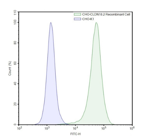
ApplicationsFlow Cytometry
TargetCLDN18
- SizePrice

Product group Antibodies
Anti-Claudin18/CLDN18 Antibody Picoband(r)A09129-1-CARRIER-FREE
ApplicationsFlow Cytometry, ImmunoFluorescence, Western Blot, ELISA, ImmunoCytoChemistry, ImmunoHistoChemistry
ReactivityHuman, Rat
TargetCLDN18
- SizePrice
Product group Antibodies
Anti-CLDN18 Antibody144-66603
ApplicationsWestern Blot
ReactivityHuman, Mouse, Rat
TargetCLDN18
- SizePrice
Product group Antibodies

Anti-Claudin-18.2 [CLDN18.2 v. 19 ]AB03747-10.0
ApplicationsFlow Cytometry
ReactivityHuman
TargetCLDN18
- SizePrice
Product group Antibodies
Anti-CLDN18 AntibodyA42555
ApplicationsWestern Blot
ReactivityHuman, Mouse
- SizePrice
Product group Antibodies
Claudin 18.2 Recombinant AntibodyBSM-60637R
ApplicationsImmunoFluorescence, ImmunoHistoChemistry, ImmunoHistoChemistry Frozen, ImmunoHistoChemistry Paraffin
ReactivityHuman, Mouse, Rat
TargetCLDN18
- SizePrice
Product group Antibodies
Cldn18 Polyclonal AntibodyCAC10906
ApplicationsWestern Blot, ELISA, ImmunoHistoChemistry
ReactivityRat
TargetCLDN18
- SizePrice
Product group Antibodies
CLDN18 AntibodyCSB-PA005498ESR1HU
ApplicationsWestern Blot, ELISA, ImmunoHistoChemistry
ReactivityHuman, Rat
TargetCLDN18
- SizePrice
Product group Antibodies

CLDN18 / Claudin 18 AntibodyLS-C409927
ApplicationsWestern Blot
ReactivityHuman, Mouse, Rat
TargetCLDN18
- SizePrice