

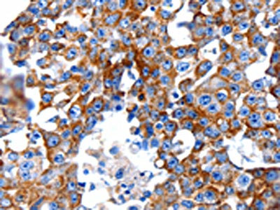
The image on the left is immunohistochemistry of paraffin-embedded Human breast cancer tissue using CSB-PA126836(CLEC7A Antibody) at dilution 1/5, on the right is treated with fusion protein. (Original magnification: x200)
 at dilution 1/5, on the right is treated with fusion protein. (Original magnification: x200)")
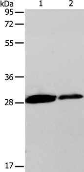
 at dilution 1/200, Secondary antibody: Goat anti rabbit IgG at 1/8000 dilution, Exposure time: 30 seconds")
The image on the left is immunohistochemistry of paraffin-embedded Human breast cancer tissue using CSB-PA126836(CLEC7A Antibody) at dilution 1/5, on the right is treated with fusion protein. (Original magnification: x200)
CLEC7A Antibody
CSB-PA126836
ApplicationsWestern Blot, ELISA, ImmunoHistoChemistry
Product group Antibodies
ReactivityHuman, Mouse
TargetCLEC7A
Overview
- SupplierCusabio
- Product NameCLEC7A Antibody
- Delivery Days Customer20
- ApplicationsWestern Blot, ELISA, ImmunoHistoChemistry
- CertificationResearch Use Only
- ClonalityPolyclonal
- ConjugateUnconjugated
- Gene ID64581
- Target nameCLEC7A
- Target descriptionC-type lectin domain containing 7A
- Target synonymsBGR, CANDF4, CD369, CLECSF12, DECTIN1, SCARE2, C-type lectin domain family 7 member A, C-type (calcium dependent, carbohydrate-recognition domain) lectin, superfamily member 12, C-type lectin superfamily member 12, DC-associated C-type lectin 1, beta-glucan receptor, dectin-1, dendritic cell-associated C-type lectin-1, lectin-like receptor 1
- HostRabbit
- IsotypeIgG
- Protein IDQ9BXN2
- Protein NameC-type lectin domain family 7 member A
- Scientific DescriptionThis gene encodes a member of the C-type lectin/C-type lectin-like domain (CTL/CTLD) superfamily. The encoded glycoprotein is a small type II membrane receptor with an extracellular C-type lectin-like domain fold and a cytoplasmic domain with an immunoreceptor tyrosine-based activation motif. It functions as a pattern-recognition receptor that recognizes a variety of beta-1,3-linked and beta-1,6-linked glucans from fungi and plants, and in this way plays a role in innate immune response. Alternate transcriptional splice variants, encoding different isoforms, have been characterized. This gene is closely linked to other CTL/CTLD superfamily members on chromosome 12p13 in the natural killer gene complex region.
- ReactivityHuman, Mouse
- Storage Instruction-20°C or -80°C
- UNSPSC41116161
Related products
Product group Antibodies
Anti-CLEC7A AntibodyA37152
ApplicationsWestern Blot, ImmunoHistoChemistry
ReactivityHuman, Mouse
- SizePrice
Product group Antibodies
Anti-CLEC7A (N-term) Antibody102-21449
ApplicationsWestern Blot, ImmunoHistoChemistry, ImmunoHistoChemistry Paraffin
TargetCLEC7A
- SizePrice

Product group Antibodies
Anti-Dectin-1/CLEC7A Antibody Picoband(r)A02731-2-CARRIER-FREE
ApplicationsFlow Cytometry, Western Blot, ELISA
ReactivityHuman
TargetCLEC7A
- SizePrice
Product group Antibodies
References
CLEC7A Polyclonal AntibodyBS-2455R
ApplicationsFlow Cytometry, Western Blot, ELISA, ImmunoHistoChemistry, ImmunoHistoChemistry Paraffin
ReactivityHuman, Mouse, Rat
TargetCLEC7A
- SizePrice
Product group Antibodies

CLEC7A / Dectin 1 AntibodyLS-C498051
ApplicationsWestern Blot
ReactivityHuman, Mouse, Rat
TargetCLEC7A
- SizePrice

Product group Antibodies
Anti-CLEC7A AntibodyHPA050229
ApplicationsWestern Blot, ImmunoCytoChemistry
ReactivityHuman
TargetCLEC7A
- SizePrice
Product group Antibodies
Dectin-1 antibody [N1C1]GTX104857
ApplicationsELISA
ReactivityHuman
TargetCLEC7A
- SizePrice
Product group Antibodies
Anti-CLEC7A AntibodyCAB9883
ApplicationsWestern Blot
ReactivityHuman
TargetCLEC7A
- SizePrice