

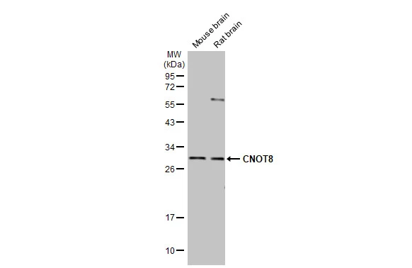
Various tissue extracts (50 μg) were separated by 12% SDS-PAGE, and the membrane was blotted with CNOT8 antibody [HL2586] (GTX639037) diluted at 1:1000. The HRP-conjugated anti-rabbit IgG antibody (GTX213110-01) was used to detect the primary antibody.
![Non-transfected (–) and transfected (+) 293T whole cell extracts were separated by 12% SDS-PAGE, and the membrane was blotted with CNOT8 antibody [HL2586] (GTX639037) diluted at 1:5000. The HRP-conjugated anti-rabbit IgG antibody (GTX213110-01) was used to detect the primary antibody.](https://www.genetex.com/upload/website/prouct_img/normal/GTX639037/GTX639037_T-45159_20230929_WB_multiple_B_23100319_368.webp "Non-transfected (–) and transfected (+) 293T whole cell extracts were separated by 12% SDS-PAGE, and the membrane was blotted with CNOT8 antibody [HL2586] (GTX639037) diluted at 1:5000. The HRP-conjugated anti-rabbit IgG antibody (GTX213110-01) was used to detect the primary antibody.")
![Various whole cell extracts (30 μg) were separated by 12% SDS-PAGE, and the membrane was blotted with CNOT8 antibody [HL2586] (GTX639037) diluted at 1:1000. The HRP-conjugated anti-rabbit IgG antibody (GTX213110-01) was used to detect the primary antibody.](https://www.genetex.com/upload/website/prouct_img/normal/GTX639037/GTX639037_45243_20231201_WB_23120519_992.webp "Various whole cell extracts (30 μg) were separated by 12% SDS-PAGE, and the membrane was blotted with CNOT8 antibody [HL2586] (GTX639037) diluted at 1:1000. The HRP-conjugated anti-rabbit IgG antibody (GTX213110-01) was used to detect the primary antibody.")
![CNOT8 antibody [HL2586] detects CNOT8 protein at cytoplasm by immunofluorescent analysis. Sample: HeLa cells were fixed in 4% paraformaldehyde at RT for 15 min. Green: CNOT8 stained by CNOT8 antibody [HL2586] (GTX639037) diluted at 1:500. Blue: Fluoroshield with DAPI (GTX30920).](https://www.genetex.com/upload/website/prouct_img/normal/GTX639037/GTX639037_T-45159_20231110_ICC_IF_23121122_472.webp "CNOT8 antibody [HL2586] detects CNOT8 protein at cytoplasm by immunofluorescent analysis. Sample: HeLa cells were fixed in 4% paraformaldehyde at RT for 15 min. Green: CNOT8 stained by CNOT8 antibody [HL2586] (GTX639037) diluted at 1:500. Blue: Fluoroshield with DAPI (GTX30920).")
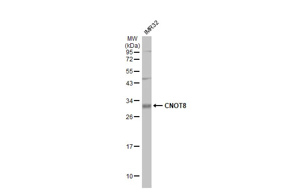
![Whole cell extract (30 μg) was separated by 12% SDS-PAGE, and the membrane was blotted with CNOT8 antibody [HL2586] (GTX639037) diluted at 1:1000. The HRP-conjugated anti-rabbit IgG antibody (GTX213110-01) was used to detect the primary antibody.](https://www.genetex.com/upload/website/prouct_img/normal/GTX639037/GTX639037_45243_20240119_WB_Z_24012217_420.webp "Whole cell extract (30 μg) was separated by 12% SDS-PAGE, and the membrane was blotted with CNOT8 antibody [HL2586] (GTX639037) diluted at 1:1000. The HRP-conjugated anti-rabbit IgG antibody (GTX213110-01) was used to detect the primary antibody.")
![Whole Japanese medaka extract (30 μg) was separated by 12% SDS-PAGE, and the membrane was blotted with CNOT8 antibody [HL2586] (GTX639037) diluted at 1:1000. The HRP-conjugated anti-rabbit IgG antibody (GTX213110-01) was used to detect the primary antibody.](https://www.genetex.com/upload/website/prouct_img/normal/GTX639037/GTX639037_45243_20250815_WB_medaka_25082121_283.webp "Whole Japanese medaka extract (30 μg) was separated by 12% SDS-PAGE, and the membrane was blotted with CNOT8 antibody [HL2586] (GTX639037) diluted at 1:1000. The HRP-conjugated anti-rabbit IgG antibody (GTX213110-01) was used to detect the primary antibody.")
Various tissue extracts (50 μg) were separated by 12% SDS-PAGE, and the membrane was blotted with CNOT8 antibody [HL2586] (GTX639037) diluted at 1:1000. The HRP-conjugated anti-rabbit IgG antibody (GTX213110-01) was used to detect the primary antibody.
CNOT8 antibody [HL2586]
GTX639037
ApplicationsImmunoFluorescence, Western Blot, ImmunoCytoChemistry
Product group Antibodies
ReactivityHuman, Mouse, Rat, Zebra Fish
TargetCNOT8
Overview
- SupplierGeneTex
- Product NameCNOT8 antibody [HL2586]
- Delivery Days Customer9
- Application Supplier NoteWB: 1:500-1:10000. *Optimal dilutions/concentrations should be determined by the researcher.Not tested in other applications.
- ApplicationsImmunoFluorescence, Western Blot, ImmunoCytoChemistry
- CertificationResearch Use Only
- ClonalityMonoclonal
- Clone IDHL2586
- Concentration1 mg/ml
- ConjugateUnconjugated
- Gene ID9337
- Target nameCNOT8
- Target descriptionCCR4-NOT transcription complex subunit 8
- Target synonymsCAF1, CALIF, Caf1b, POP2, hCAF1, CCR4-NOT transcription complex subunit 8, CAF1-like protein, CAF2, CALIFp, CCR4-associated factor 8, PGK promoter directed over production
- HostRabbit
- IsotypeIgG
- Protein IDQ9UFF9
- Protein NameCCR4-NOT transcription complex subunit 8
- Scientific DescriptionEnables poly(A)-specific ribonuclease activity. Involved in exonucleolytic catabolism of deadenylated mRNA and positive regulation of cell population proliferation. Located in nucleus. Part of CCR4-NOT complex. [provided by Alliance of Genome Resources, Apr 2022]
- ReactivityHuman, Mouse, Rat, Zebra Fish
- Storage Instruction-20°C or -80°C,2°C to 8°C
- UNSPSC41116161
Datasheet
Related products

Product group Antibodies
Anti-CNOT8 Antibody (C-term)A05401-1
ApplicationsFlow Cytometry, ImmunoFluorescence, Western Blot, ImmunoHistoChemistry, ImmunoHistoChemistry Paraffin
ReactivityHuman, Mouse
TargetCNOT8
- SizePrice

Product group Antibodies
Anti-CNOT8 AntibodyA16055
ApplicationsWestern Blot
ReactivityHuman, Mouse
- SizePrice
Product group Antibodies
Anti-CNOT8 Antibody144-08058
ApplicationsWestern Blot, ImmunoHistoChemistry
ReactivityHuman, Mouse, Rat
TargetCNOT8
- SizePrice
Product group Antibodies

CNOT8 AntibodyLS-C747645
ApplicationsWestern Blot
ReactivityHuman, Mouse, Rat
TargetCNOT8
- SizePrice
Product group Antibodies
CNOT8 Polyclonal AntibodyBS-9388R
ApplicationsImmunoFluorescence, Western Blot, ImmunoHistoChemistry, ImmunoHistoChemistry Paraffin
ReactivityChicken, Human, Mouse, Rat
TargetCNOT8
- SizePrice
Product group Antibodies
CNOT8 AntibodyCSB-PA871398ESR2HU
ApplicationsELISA, ImmunoHistoChemistry
ReactivityHuman
TargetCNOT8
- SizePrice
Product group Antibodies
CNOT8 antibody, C-termGTX81874
ApplicationsFlow Cytometry, ImmunoFluorescence, Western Blot, ImmunoCytoChemistry, ImmunoHistoChemistry, ImmunoHistoChemistry Paraffin
ReactivityHuman
TargetCNOT8
- SizePrice

Product group Antibodies
Anti-CNOT8 AntibodyHPA051398
ApplicationsImmunoHistoChemistry
ReactivityHuman
TargetCNOT8
- SizePrice
Product group Antibodies
CNOT8 antibodyGTX105674
ApplicationsImmunoFluorescence, Western Blot, ImmunoCytoChemistry
ReactivityHuman
TargetCNOT8
- SizePrice
Product group Antibodies
CNOT8 antibodyGTX64486
ApplicationsWestern Blot, ImmunoHistoChemistry, ImmunoHistoChemistry Paraffin
ReactivityHuman, Mouse
TargetCNOT8
- SizePrice