

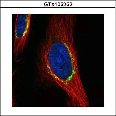
Confocal immunofluorescence analysis (Olympus FV10i) of paraformaldehyde-fixed HeLa, using COPD(GTX103252) antibody (green) at 1:500 dilution. Alpha-tubulin filaments were labeled with GTX11304 (red) at 1:2500.
 A:H1299 B:Hep G2(GTX27900) 7.5% SDS PAGE GTX103252 diluted at 1:500")
 diluted at 1:500.
Antigen Retrieval: Citrate buffer, pH 6.0, 10 min")
 and transfected (+) 293T whole cell extracts (30 μg) were separated by 7.5% SDS-PAGE, and the membrane was blotted with COPD antibody (GTX103252) diluted at 1:2000.")
![COPD antibody detects COPD protein by immunohistochemical analysis. Sample: Frozen-sectioned mouse mouse cerebellum. Green: COPD stained by COPD antibody (GTX103252) diluted at 1:250. Red: NF-H, stained by NF-H antibody [GT114] (GTX634289) diluted at 1:500. Blue: Fluoroshield with DAPI (GTX30920).
Antigen Retrieval: Citrate buffer, pH 6.0, 10 min](https://www.genetex.com/upload/website/prouct_img/normal/GTX103252/GTX103252_40240_20180530_IHC-Fr_M_2_w_23060119_381.webp "COPD antibody detects COPD protein by immunohistochemical analysis. Sample: Frozen-sectioned mouse mouse cerebellum. Green: COPD stained by COPD antibody (GTX103252) diluted at 1:250. Red: NF-H, stained by NF-H antibody [GT114] (GTX634289) diluted at 1:500. Blue: Fluoroshield with DAPI (GTX30920).
Antigen Retrieval: Citrate buffer, pH 6.0, 10 min")
Confocal immunofluorescence analysis (Olympus FV10i) of paraformaldehyde-fixed HeLa, using COPD(GTX103252) antibody (green) at 1:500 dilution. Alpha-tubulin filaments were labeled with GTX11304 (red) at 1:2500.
COPD antibody
GTX103252
ApplicationsImmunoFluorescence, Western Blot, ImmunoCytoChemistry, ImmunoHistoChemistry, ImmunoHistoChemistry Frozen, ImmunoHistoChemistry Paraffin
Product group Antibodies
ReactivityHuman, Mouse
TargetARCN1
Overview
- SupplierGeneTex
- Product NameCOPD antibody
- Delivery Days Customer9
- Application Supplier NoteWB: 1:500-1:3000. ICC/IF: 1:100-1:1000. IHC-P: 1:100-1:1000. IHC-Fr: 1:100-1:1000. *Optimal dilutions/concentrations should be determined by the researcher.Not tested in other applications.
- ApplicationsImmunoFluorescence, Western Blot, ImmunoCytoChemistry, ImmunoHistoChemistry, ImmunoHistoChemistry Frozen, ImmunoHistoChemistry Paraffin
- CertificationResearch Use Only
- ClonalityPolyclonal
- Concentration0.21 mg/ml
- ConjugateUnconjugated
- Gene ID372
- Target nameARCN1
- Target descriptionarchain 1 coat protein complex I subunit delta
- Target synonymsCOPD, SRMMD, SSMG, coatomer subunit delta, COPI coat complex subunit delta, archain vesicle transport protein 1, coatomer delta subunit, coatomer protein complex, subunit delta, coatomer protein delta-COP, delta-COP, delta-coat protein
- HostRabbit
- IsotypeIgG
- Protein IDP48444
- Protein NameCoatomer subunit delta
- Scientific DescriptionThis gene maps in a region, which include the mixed lineage leukemia and Friend leukemia virus integration 1 genes, where multiple disease-associated chromosome translocations occur. It is an intracellular protein. Archain sequences are well conserved among eukaryotes and this protein may play a fundamental role in eukaryotic cell biology. It has similarities to heat shock proteins and clathrin-associated proteins, and may be involved in vesicle structure or trafficking. [provided by RefSeq]
- ReactivityHuman, Mouse
- Storage Instruction-20°C or -80°C,2°C to 8°C
- UNSPSC41116161
Datasheet
Related products

Product group Antibodies
Anti-COPD AntibodyA39891
ApplicationsImmunoFluorescence, Western Blot
ReactivityHuman
- SizePrice
Product group Antibodies

COPD / ARCN1 AntibodyLS-C831213
ApplicationsELISA, ImmunoHistoChemistry
ReactivityHuman, Mouse, Rat
TargetARCN1
- SizePrice

Product group Antibodies
Anti-ARCN1 AntibodyHPA037573
ApplicationsWestern Blot, ImmunoCytoChemistry, ImmunoHistoChemistry
ReactivityHuman, Mouse, Rat
TargetARCN1
- SizePrice
![COPD antibody [GT189] detects COPD protein by western blot analysis. A. 30 μg 293T whole cell lysate/extract B. 30 μg A431 whole cell lysate/extract C. 30 μg HeLa whole cell lysate/extract D. 30 μg HepG2 whole cell lysate/extract 7.5 % SDS-PAGE COPD antibody [GT189] (GTX630561) dilution: 1:10000](https://www.genetex.com/upload/website/prouct_img/normal/GTX630561/GTX630561_41589_WB_w_23061202_678.webp)
Product group Antibodies
COPD antibody [GT189]GTX630561
ApplicationsWestern Blot, ImmunoHistoChemistry, ImmunoHistoChemistry Frozen, ImmunoHistoChemistry Paraffin
ReactivityHuman, Mouse, Rat
TargetARCN1
- SizePrice
![COPD antibody [GT1318] detects COPD protein by western blot analysis. A. 30 μg PC-12 whole cell lysate/extract B. 30 μg Rat2 whole cell lysate/extract 7.5 % SDS-PAGE COPD antibody [GT1318] (GTX630562) dilution: 1:1000](https://www.genetex.com/upload/website/prouct_img/normal/GTX630562/GTX630562_41589_WB_R_w_23061202_798.webp)
Product group Antibodies
COPD antibody [GT1318]GTX630562
ApplicationsImmunoFluorescence, Western Blot, ImmunoCytoChemistry, ImmunoHistoChemistry, ImmunoHistoChemistry Frozen, ImmunoHistoChemistry Paraffin
ReactivityHuman, Mouse, Rat
TargetARCN1
- SizePrice
Product group Antibodies
Anti-ARCN1 Antibody144-60876
ApplicationsWestern Blot
ReactivityHuman, Mouse
TargetARCN1
- SizePrice