

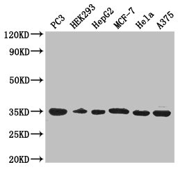
Western Blot Positive WB detected in: PC-3 whole cell lysate, HEK293 whole cell lysate, HepG2 whole cell lysate, MCF-7 whole cell lysate, Hela whole cell lysate, A375 whole cell lysate All lanes: COPE antibody at 3microg/ml Secondary Goat polyclonal to rabbit IgG at 1/50000 dilution Predicted band size: 35, 29 kDa Observed band size: 35 kDa

Western Blot Positive WB detected in: PC-3 whole cell lysate, HEK293 whole cell lysate, HepG2 whole cell lysate, MCF-7 whole cell lysate, Hela whole cell lysate, A375 whole cell lysate All lanes: COPE antibody at 3microg/ml Secondary Goat polyclonal to rabbit IgG at 1/50000 dilution Predicted band size: 35, 29 kDa Observed band size: 35 kDa
COPE Antibody
CSB-PA03669A0RB
ApplicationsWestern Blot, ELISA, ImmunoHistoChemistry
Product group Antibodies
ReactivityHuman
TargetCOPE
Overview
- SupplierCusabio
- Product NameCOPE Antibody
- Delivery Days Customer20
- ApplicationsWestern Blot, ELISA, ImmunoHistoChemistry
- CertificationResearch Use Only
- ClonalityPolyclonal
- ConjugateUnconjugated
- Gene ID11316
- Target nameCOPE
- Target descriptioncoat protein complex I subunit epsilon
- Target synonymsepsilon-COP, coatomer subunit epsilon, COPI coat complex subunit epsilon, coatomer epsilon subunit, coatomer protein complex subunit epsilon, epsilon coat protein
- HostRabbit
- IsotypeIgG
- Protein IDO14579
- Protein NameCoatomer subunit epsilon
- Scientific DescriptionThe coatomer is a cytosolic protein complex that binds to dilysine motifs and reversibly associates with Golgi non-clathrin-coated vesicles, which further mediate biosynthetic protein transport from the ER, via the Golgi up to the trans Golgi network. The coatomer complex is required for budding from Golgi membranes, and is essential for the retrograde Golgi-to-ER transport of dilysine-tagged proteins. In mammals, the coatomer can only be recruited by membranes associated with ADP-ribosylation factors (ARFs), which are small GTP-binding proteins; the complex also influences the Golgi structural integrity, as well as the processing, activity, and endocytic recycling of LDL receptors.
- ReactivityHuman
- Storage Instruction-20°C or -80°C
- UNSPSC41116161
Related products
Product group Antibodies
Cope Polyclonal AntibodyCAC09032
ApplicationsWestern Blot, ELISA, ImmunoHistoChemistry
TargetCOPE
- SizePrice
Product group Antibodies
Anti-COPE AntibodyA11226
ApplicationsWestern Blot
ReactivityHuman, Mouse, Rat
- SizePrice
Product group Antibodies
Anti-COPE Antibody144-10047
ApplicationsWestern Blot
ReactivityHuman, Mouse
TargetCOPE
- SizePrice
Product group Antibodies

COPE AntibodyLS-C832104
ApplicationsWestern Blot, ELISA, ImmunoHistoChemistry
ReactivityHuman, Mouse
TargetCOPE
- SizePrice

Product group Antibodies
Anti-COPE AntibodyHPA041605
ApplicationsImmunoCytoChemistry, ImmunoHistoChemistry
ReactivityHuman
TargetCOPE
- SizePrice
Product group Antibodies
Anti-COPE Antibody Picoband(r)A04544-CARRIER-FREE
ApplicationsFlow Cytometry, ImmunoFluorescence, Western Blot, ImmunoCytoChemistry, ImmunoHistoChemistry, ImmunoHistoChemistry Frozen
ReactivityHuman, Mouse, Rat
TargetCOPE
- SizePrice
Product group Antibodies
COPE antibodyGTX64942
ApplicationsWestern Blot
ReactivityHuman, Mouse
TargetCOPE
- SizePrice
Product group Antibodies
COPE Monoclonal AntibodyBSM-60452M
ApplicationsImmunoFluorescence, Western Blot, ImmunoCytoChemistry
ReactivityHuman
TargetCOPE
- SizePrice
Product group Antibodies
COPEY058504
ApplicationsWestern Blot, ImmunoHistoChemistry
ReactivityHuman
- SizePrice