

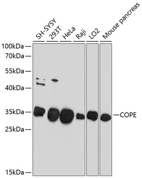
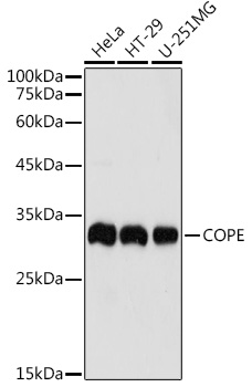
WB analysis of various sample lysates using GTX64942 COPE antibody. Dilution : 1:1000 Loading : 25μg per lane
WB analysis of various sample lysates using GTX64942 COPE antibody. Dilution : 1:1000 Loading : 25μg per lane
COPE antibody
GTX64942
ApplicationsWestern Blot
Product group Antibodies
ReactivityHuman, Mouse
TargetCOPE
Overview
- SupplierGeneTex
- Product NameCOPE antibody
- Delivery Days Customer9
- Application Supplier NoteWB: 1:500 - 1:2000. *Optimal dilutions/concentrations should be determined by the researcher.Not tested in other applications.
- ApplicationsWestern Blot
- CertificationResearch Use Only
- ClonalityPolyclonal
- ConjugateUnconjugated
- Gene ID11316
- Target nameCOPE
- Target descriptioncoat protein complex I subunit epsilon
- Target synonymsepsilon-COP, coatomer subunit epsilon, COPI coat complex subunit epsilon, coatomer epsilon subunit, coatomer protein complex subunit epsilon, epsilon coat protein
- HostRabbit
- IsotypeIgG
- Protein IDO14579
- Protein NameCoatomer subunit epsilon
- Scientific DescriptionThe product of this gene is an epsilon subunit of coatomer protein complex. Coatomer is a cytosolic protein complex that binds to dilysine motifs and reversibly associates with Golgi non-clathrin-coated vesicles. It is required for budding from Golgi membranes, and is essential for the retrograde Golgi-to-ER transport of dilysine-tagged proteins. Coatomer complex consists of at least the alpha, beta, beta, gamma, delta, epsilon and zeta subunits. Alternatively spliced transcript variants encoding different isoforms have been identified. [provided by RefSeq, Jul 2008]
- ReactivityHuman, Mouse
- Storage Instruction-20°C or -80°C,2°C to 8°C
- UNSPSC41116161
Datasheet
Related products
Product group Antibodies
Cope Polyclonal AntibodyCAC09032
ApplicationsWestern Blot, ELISA, ImmunoHistoChemistry
TargetCOPE
- SizePrice
Product group Antibodies
Anti-COPE AntibodyA11226
ApplicationsWestern Blot
ReactivityHuman, Mouse, Rat
- SizePrice
Product group Antibodies
Anti-COPE Antibody144-10047
ApplicationsWestern Blot
ReactivityHuman, Mouse
TargetCOPE
- SizePrice
Product group Antibodies

COPE AntibodyLS-C832104
ApplicationsWestern Blot, ELISA, ImmunoHistoChemistry
ReactivityHuman, Mouse
TargetCOPE
- SizePrice
Product group Antibodies
COPE AntibodyCSB-PA03669A0RB
ApplicationsWestern Blot, ELISA, ImmunoHistoChemistry
ReactivityHuman
TargetCOPE
- SizePrice

Product group Antibodies
Anti-COPE AntibodyHPA041605
ApplicationsImmunoCytoChemistry, ImmunoHistoChemistry
ReactivityHuman
TargetCOPE
- SizePrice
Product group Antibodies
Anti-COPE Antibody Picoband(r)A04544-CARRIER-FREE
ApplicationsFlow Cytometry, ImmunoFluorescence, Western Blot, ImmunoCytoChemistry, ImmunoHistoChemistry, ImmunoHistoChemistry Frozen
ReactivityHuman, Mouse, Rat
TargetCOPE
- SizePrice
Product group Antibodies
COPE Monoclonal AntibodyBSM-60452M
ApplicationsImmunoFluorescence, Western Blot, ImmunoCytoChemistry
ReactivityHuman
TargetCOPE
- SizePrice
Product group Antibodies
COPEY058504
ApplicationsWestern Blot, ImmunoHistoChemistry
ReactivityHuman
- SizePrice