

COT antibody
70R-30667
Product group Antibodies
Overview
- SupplierBiosynth
- Product NameCOT antibody
- Delivery Days Customer11
- CertificationResearch Use Only
- UNSPSC41116161
Related products
Product group Antibodies
Anti-MAP3K8 AntibodyA95620
ApplicationsWestern Blot, ELISA, ImmunoHistoChemistry
ReactivityHuman, Mouse, Rat
- SizePrice
Product group Antibodies

MAP3K8 (Phospho-Ser400) AntibodyABX012608
ApplicationsELISA, ImmunoHistoChemistry
- SizePrice
Product group Antibodies
ApplicationsWestern Blot, ImmunoHistoChemistry, ImmunoHistoChemistry Paraffin
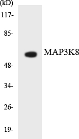
TargetMAP3K8
- SizePrice
Product group Antibodies
MAP3K8 Polyclonal AntibodyBS-21629R
ApplicationsWestern Blot
ReactivityBovine, Canine, Equine, Human, Mouse, Porcine, Rabbit, Rat, Sheep
TargetMAP3K8
- SizePrice
Product group Antibodies
MAP3K8 AntibodyCSB-PA001760
ApplicationsImmunoFluorescence, Western Blot, ELISA, ImmunoHistoChemistry
ReactivityHuman, Mouse, Rat
TargetMAP3K8
- SizePrice
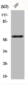
![Various whole cell extracts (30 μg) were separated by 7.5% SDS-PAGE, and the membrane was blotted with MAP3K8 antibody [N3C3] (GTX102711) diluted at 1:1000. The HRP-conjugated anti-rabbit IgG antibody (GTX213110-01) was used to detect the primary antibody. Corresponding RNA expression data for the same cell lines are based on Human Protein Atlas program.](https://www.genetex.com/upload/website/prouct_img/normal/GTX102711/GTX102711_44916_20230331_WB_TPM_watermark_23041023_238.webp)
Product group Antibodies
MAP3K8 antibody [N3C3]GTX102711
ApplicationsImmunoFluorescence, Western Blot, ImmunoCytoChemistry, ImmunoHistoChemistry, ImmunoHistoChemistry Paraffin
ReactivityHuman, Mouse
TargetMAP3K8
- SizePrice
Product group Antibodies

MAP3K8 / TPL2 Antibody (Internal)LS-C358799
ApplicationsImmunoFluorescence, Western Blot, ImmunoCytoChemistry, ImmunoHistoChemistry, ImmunoHistoChemistry Paraffin
ReactivityBovine, Human, Monkey, Mouse, Rat
TargetMAP3K8
- SizePrice

Product group Antibodies
Anti-MAP3K8 AntibodyHPA017962
ApplicationsWestern Blot, ImmunoCytoChemistry, ImmunoHistoChemistry
ReactivityHuman
TargetMAP3K8
- SizePrice

Product group Antibodies
Anti-MAP3K8 Antibody Picoband(r)PB10007-CARRIER-FREE
ApplicationsFlow Cytometry, ImmunoFluorescence, Western Blot, ImmunoCytoChemistry, ImmunoHistoChemistry
ReactivityHuman, Rat
TargetMAP3K8
- SizePrice