

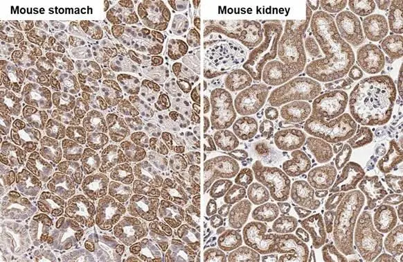
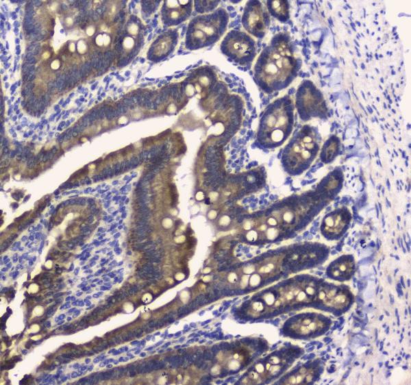
COX4 antibody [HL2264] detects COX4 protein by immunohistochemical analysis. Sample: Paraffin-embedded mouse tissues. COX4 stained by COX4 antibody [HL2264] (GTX638316) diluted at 1:100. Antigen Retrieval: Citrate buffer, pH 6.0, 15 min
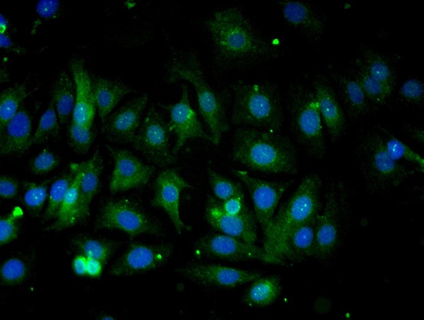
![COX4 antibody [HL2264] detects COX4 protein at mitochondria by immunofluorescent analysis. Sample: HeLa cells were fixed in ice-cold MeOH for 5 min. Green: COX4 stained by COX4 antibody [HL2264] (GTX638316) diluted at 1:500. Red: alpha Tubulin, a cytoskeleton marker, stained by alpha Tubulin antibody [GT114] (GTX628802) diluted at 1:1000. Blue: Fluoroshield with DAPI (GTX30920).](https://www.genetex.com/upload/website/prouct_img/normal/GTX638316/GTX638316_T-44970_20230421_ICC_IF_23050918_382.webp "COX4 antibody [HL2264] detects COX4 protein at mitochondria by immunofluorescent analysis. Sample: HeLa cells were fixed in ice-cold MeOH for 5 min. Green: COX4 stained by COX4 antibody [HL2264] (GTX638316) diluted at 1:500. Red: alpha Tubulin, a cytoskeleton marker, stained by alpha Tubulin antibody [GT114] (GTX628802) diluted at 1:1000. Blue: Fluoroshield with DAPI (GTX30920).")
![Various tissue extracts (50 μg) were separated by 15% SDS-PAGE, and the membrane was blotted with COX4 antibody [HL2264] (GTX638316) diluted at 1:1000. The HRP-conjugated anti-rabbit IgG antibody (GTX213110-01) was used to detect the primary antibody.](https://www.genetex.com/upload/website/prouct_img/normal/GTX638316/GTX638316_45033_20230519_WB_M_R_23053001_150.webp "Various tissue extracts (50 μg) were separated by 15% SDS-PAGE, and the membrane was blotted with COX4 antibody [HL2264] (GTX638316) diluted at 1:1000. The HRP-conjugated anti-rabbit IgG antibody (GTX213110-01) was used to detect the primary antibody.")
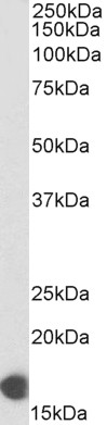
![Rat tissue extract (50 μg) was separated by 15% SDS-PAGE, and the membrane was blotted with COX4 antibody [HL2264] (GTX638316) diluted at 1:1000. The HRP-conjugated anti-rabbit IgG antibody (GTX213110-01) was used to detect the primary antibody, and the signal was developed with Trident ECL plus-Enhanced.](https://www.genetex.com/upload/website/prouct_img/normal/GTX638316/GTX638316_45033_20230519_WB_R_stomach_23053001_258.webp "Rat tissue extract (50 μg) was separated by 15% SDS-PAGE, and the membrane was blotted with COX4 antibody [HL2264] (GTX638316) diluted at 1:1000. The HRP-conjugated anti-rabbit IgG antibody (GTX213110-01) was used to detect the primary antibody, and the signal was developed with Trident ECL plus-Enhanced.")
![Whole cell extract (30 μg) was separated by 15% SDS-PAGE, and the membrane was blotted with COX4 antibody [HL2264] (GTX638316) diluted at 1:1000. The HRP-conjugated anti-rabbit IgG antibody (GTX213110-01) was used to detect the primary antibody, and the signal was developed with Trident femto Western HRP Substrate.](https://www.genetex.com/upload/website/prouct_img/normal/GTX638316/GTX638316_45033_20230526_WB_M_23053001_859.webp "Whole cell extract (30 μg) was separated by 15% SDS-PAGE, and the membrane was blotted with COX4 antibody [HL2264] (GTX638316) diluted at 1:1000. The HRP-conjugated anti-rabbit IgG antibody (GTX213110-01) was used to detect the primary antibody, and the signal was developed with Trident femto Western HRP Substrate.")
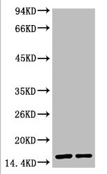
![Whole zebrafish extract (30 μg) was separated by 15% SDS-PAGE, and the membrane was blotted with COX4 antibody [HL2264] (GTX638316) diluted at 1:1000. The HRP-conjugated anti-rabbit IgG antibody (GTX213110-01) was used to detect the primary antibody, and the signal was developed with Trident ECL plus-Enhanced.](https://www.genetex.com/upload/website/prouct_img/normal/GTX638316/GTX638316_45033_20230609_WB_Z_23061400_250.webp "Whole zebrafish extract (30 μg) was separated by 15% SDS-PAGE, and the membrane was blotted with COX4 antibody [HL2264] (GTX638316) diluted at 1:1000. The HRP-conjugated anti-rabbit IgG antibody (GTX213110-01) was used to detect the primary antibody, and the signal was developed with Trident ECL plus-Enhanced.")
![Non-transfected (–) and transfected (+) 293T whole cell extracts (30 μg) were separated by 15% SDS-PAGE, and the membrane was blotted with COX4 antibody [HL2264] (GTX638316) diluted at 1:1000. The HRP-conjugated anti-rabbit IgG antibody (GTX213110-01) was used to detect the primary antibody.](https://www.genetex.com/upload/website/prouct_img/normal/GTX638316/GTX638316_45033_20230714_WB_shRNA_watermark_23071822_334.webp "Non-transfected (–) and transfected (+) 293T whole cell extracts (30 μg) were separated by 15% SDS-PAGE, and the membrane was blotted with COX4 antibody [HL2264] (GTX638316) diluted at 1:1000. The HRP-conjugated anti-rabbit IgG antibody (GTX213110-01) was used to detect the primary antibody.")
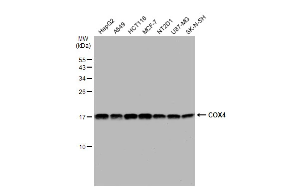
![Various whole cell extracts (30 μg) were separated by 12% SDS-PAGE, and the membrane was blotted with COX4 antibody [HL2264] (GTX638316) diluted at 1:1000. The HRP-conjugated anti-rabbit IgG antibody (GTX213110-01) was used to detect the primary antibody.](https://www.genetex.com/upload/website/prouct_img/normal/GTX638316/GTX638316_45033_20241004_WB_24100900_441.webp "Various whole cell extracts (30 μg) were separated by 12% SDS-PAGE, and the membrane was blotted with COX4 antibody [HL2264] (GTX638316) diluted at 1:1000. The HRP-conjugated anti-rabbit IgG antibody (GTX213110-01) was used to detect the primary antibody.")
COX4 antibody [HL2264] detects COX4 protein by immunohistochemical analysis. Sample: Paraffin-embedded mouse tissues. COX4 stained by COX4 antibody [HL2264] (GTX638316) diluted at 1:100. Antigen Retrieval: Citrate buffer, pH 6.0, 15 min
COX4 antibody [HL2264]
GTX638316
ApplicationsImmunoFluorescence, Western Blot, ImmunoCytoChemistry, ImmunoHistoChemistry, ImmunoHistoChemistry Paraffin
Product group Antibodies
ReactivityHuman, Mouse, Rat, Zebra Fish
TargetCOX4I1
Overview
- SupplierGeneTex
- Product NameCOX4 antibody [HL2264]
- Delivery Days Customer9
- Application Supplier NoteWB: 1:500-1:3000. *Optimal dilutions/concentrations should be determined by the researcher.Not tested in other applications.
- ApplicationsImmunoFluorescence, Western Blot, ImmunoCytoChemistry, ImmunoHistoChemistry, ImmunoHistoChemistry Paraffin
- CertificationResearch Use Only
- ClonalityMonoclonal
- Clone IDHL2264
- Concentration1 mg/ml
- ConjugateUnconjugated
- Gene ID1327
- Target nameCOX4I1
- Target descriptioncytochrome c oxidase subunit 4I1
- Target synonymsCOX IV-1, COX4, COX4-1, COXIV, COXIV-1, MC4DN16, cytochrome c oxidase subunit 4 isoform 1, mitochondrial, cytochrome c oxidase polypeptide IV, cytochrome c oxidase subunit IV
- HostRabbit
- IsotypeIgG
- Protein IDP13073
- Protein NameCytochrome c oxidase subunit 4 isoform 1, mitochondrial
- Scientific DescriptionCytochrome c oxidase (COX) is the terminal enzyme of the mitochondrial respiratory chain. It is a multi-subunit enzyme complex that couples the transfer of electrons from cytochrome c to molecular oxygen and contributes to a proton electrochemical gradient across the inner mitochondrial membrane. The complex consists of 13 mitochondrial- and nuclear-encoded subunits. The mitochondrially-encoded subunits perform the electron transfer and proton pumping activities. The functions of the nuclear-encoded subunits are unknown but they may play a role in the regulation and assembly of the complex. This gene encodes the nuclear-encoded subunit IV isoform 1 of the human mitochondrial respiratory chain enzyme. It is located at the 3 of the NOC4 (neighbor of COX4) gene in a head-to-head orientation, and shares a promoter with it. Pseudogenes related to this gene are located on chromosomes 13 and 14. Alternative splicing results in multiple transcript variants encoding different isoforms. [provided by RefSeq, Jan 2016]
- ReactivityHuman, Mouse, Rat, Zebra Fish
- Storage Instruction-20°C or -80°C,2°C to 8°C
- UNSPSC41116161
Datasheet
Related products
Product group Antibodies
ApplicationsWestern Blot, ELISA
ReactivityHuman
- SizePrice
Product group Antibodies
Anti-COX IV/COX4I1 Antibody Picoband(r)A05442-1-CARRIER-FREE
ApplicationsFlow Cytometry, ImmunoFluorescence, Western Blot, ELISA, ImmunoCytoChemistry, ImmunoHistoChemistry
ReactivityHuman, Mouse, Rat
TargetCOX4I1
- SizePrice

Product group Antibodies
Anti-COX4I1 AntibodyAMAB91171
ApplicationsWestern Blot, ImmunoCytoChemistry, ImmunoHistoChemistry
ReactivityHuman, Mouse, Rat
TargetCOX4I1
- SizePrice
Product group Antibodies

COXIV / COX4 Antibody (clone 2D4)LS-C765752
ApplicationsImmunoFluorescence, Western Blot, ImmunoHistoChemistry, ImmunoHistoChemistry Paraffin
ReactivityHuman, Mouse, Rat
TargetCOX4I1
- SizePrice
Product group Antibodies
References
COX4I1 Polyclonal AntibodyBS-10257R
ApplicationsFlow Cytometry, ImmunoFluorescence, Western Blot, ELISA, ImmunoCytoChemistry, ImmunoHistoChemistry, ImmunoHistoChemistry Frozen, ImmunoHistoChemistry Paraffin
ReactivityBovine, Canine, Equine, Human, Mouse, Porcine, Rat
TargetCOX4I1
- SizePrice
Product group Antibodies
COX4I1 Monoclonal AntibodyCSB-MA000188
ApplicationsWestern Blot, ELISA
ReactivityHuman, Mouse, Rat
TargetCOX4I1
- SizePrice
Product group Antibodies

Goat anti-COX4I1, BiotinylatedEB07914-B
ApplicationsWestern Blot, ELISA, ImmunoHistoChemistry
ReactivityHuman
TargetCOX4I1
- SizePrice
Product group Antibodies
Cox4I1 Polyclonal AntibodyCAC07075
ApplicationsWestern Blot, ELISA, ImmunoHistoChemistry
ReactivityRat, Zebra Fish
TargetCOX4I1
- SizePrice
Product group Antibodies
COX4 antibodyGTX101499
ApplicationsImmunoFluorescence, ImmunoPrecipitation, Western Blot, ImmunoCytoChemistry, ImmunoHistoChemistry, ImmunoHistoChemistry Paraffin
ReactivityHuman, Mouse
TargetCOX4I1
- SizePrice