

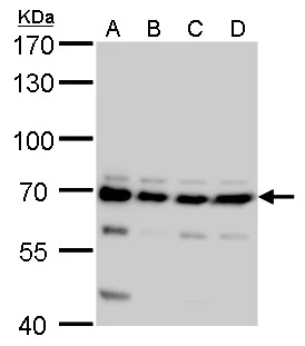
CPSF6 antibody detects CPSF6 protein by Western blot analysis. A. 30 μg 293T B. 30 μg A431 C. 30 μg HeLa D. 30 μg HepG2 7.5 % SDS-PAGE CPSF6 antibody (GTX115537) dilution: 1:5000
 dilution: 1:500.
Antigen Retrieval: Trilogy? (EDTA based, pH 8.0) buffer, 15min")
 dilution: 1:500.
Antigen Retrieval: Trilogy? (EDTA based, pH 8.0) buffer, 15min")
 diluted at 1:500. Blue: Hoechst 33342 staining.")
CPSF6 antibody detects CPSF6 protein by Western blot analysis. A. 30 μg 293T B. 30 μg A431 C. 30 μg HeLa D. 30 μg HepG2 7.5 % SDS-PAGE CPSF6 antibody (GTX115537) dilution: 1:5000
CPSF6 antibody
GTX115537
ApplicationsImmunoFluorescence, Western Blot, ImmunoCytoChemistry, ImmunoHistoChemistry, ImmunoHistoChemistry Paraffin
Product group Antibodies
ReactivityHuman, Mouse, Rat
TargetCPSF6
Overview
- SupplierGeneTex
- Product NameCPSF6 antibody
- Delivery Days Customer9
- Application Supplier NoteWB: 1:1000-1:10000. ICC/IF: 1:100-1:1000. IHC-P: 1:100-1:1000. *Optimal dilutions/concentrations should be determined by the researcher.Not tested in other applications.
- ApplicationsImmunoFluorescence, Western Blot, ImmunoCytoChemistry, ImmunoHistoChemistry, ImmunoHistoChemistry Paraffin
- CertificationResearch Use Only
- ClonalityPolyclonal
- Concentration1 mg/ml
- ConjugateUnconjugated
- Gene ID11052
- Target nameCPSF6
- Target descriptioncleavage and polyadenylation specific factor 6
- Target synonymsCFIM, CFIM68, CFIM72, HPBRII-4, HPBRII-7, cleavage and polyadenylation specificity factor subunit 6, CPSF 68 kDa subunit, cleavage and polyadenylation specific factor 6, 68kDa, cleavage and polyadenylation specificity factor 68 kDa subunit, cleavage factor Im complex 68 kDa subunit, pre-mRNA cleavage factor I, 68kD subunit, pre-mRNA cleavage factor Im (68kD), pre-mRNA cleavage factor Im 68 kDa subunit, protein HPBRII-4/7
- HostRabbit
- IsotypeIgG
- Protein IDQ16630
- Protein NameCleavage and polyadenylation specificity factor subunit 6
- Scientific DescriptionThe protein encoded by this gene is one subunit of a cleavage factor required for 3 RNA cleavage and polyadenylation processing. The interaction of the protein with the RNA is one of the earliest steps in the assembly of the 3 end processing complex and facilitates the recruitment of other processing factors. The cleavage factor complex is composed of four polypeptides. This gene encodes the 68kD subunit. It has a domain organization reminiscent of spliceosomal proteins. [provided by RefSeq]
- ReactivityHuman, Mouse, Rat
- Storage Instruction-20°C or -80°C,2°C to 8°C
- UNSPSC12352203
Datasheet
Related products
Product group Antibodies
Anti-CPSF6 Antibody144-05963
ApplicationsWestern Blot
ReactivityHuman, Mouse, Rat
TargetCPSF6
- SizePrice
Product group Antibodies
CPSF6 Polyclonal AntibodyCAC14997
ApplicationsImmunoFluorescence, Western Blot, ELISA, ImmunoHistoChemistry
ReactivityMouse
TargetCPSF6
- SizePrice
Product group Antibodies
Anti-CPSF6 AntibodyA15061
ApplicationsImmunoFluorescence, ImmunoPrecipitation, Western Blot, ImmunoCytoChemistry, ImmunoHistoChemistry
ReactivityHuman, Mouse, Rat
- SizePrice
Product group Antibodies
CPSF6 antibody, InternalGTX47333
ApplicationsImmunoFluorescence, Western Blot, ImmunoCytoChemistry, ImmunoHistoChemistry, ImmunoHistoChemistry Paraffin
ReactivityHuman
TargetCPSF6
- SizePrice

Product group Antibodies
Anti-CPSF6 AntibodyHPA039973
ApplicationsWestern Blot, ImmunoCytoChemistry, ImmunoHistoChemistry
ReactivityHuman, Mouse, Rat
TargetCPSF6
- SizePrice
Product group Antibodies
CPSF6 AntibodyCSB-PA621966LA01HU
ApplicationsImmunoFluorescence, Western Blot, ELISA, ImmunoHistoChemistry
ReactivityHuman, Mouse
TargetCPSF6
- SizePrice
Product group Antibodies

CPSF6 AntibodyLS-C498021
ApplicationsWestern Blot
ReactivityHuman, Mouse, Rat
TargetCPSF6
- SizePrice

Product group Antibodies
Anti-CPSF6 Antibody Picoband(r)A04551-1-CARRIER-FREE
ApplicationsImmunoFluorescence, Western Blot, ELISA, ImmunoCytoChemistry, ImmunoHistoChemistry
ReactivityHuman, Monkey, Mouse, Rat
TargetCPSF6
- SizePrice