

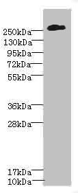
Western blot All lanes: CSPG4 antibody at 2.51microg/ml + A375 whole cell lysate Secondary Goat polyclonal to rabbit IgG at 1/10000 dilution Predicted band size: 251 kDa Observed band size: 251 kDa

Western blot All lanes: CSPG4 antibody at 2.51microg/ml + A375 whole cell lysate Secondary Goat polyclonal to rabbit IgG at 1/10000 dilution Predicted band size: 251 kDa Observed band size: 251 kDa
CSPG4 Antibody
CSB-PA006076ESR1HU
ApplicationsWestern Blot, ELISA, ImmunoHistoChemistry
Product group Antibodies
ReactivityHuman
TargetCSPG4
Overview
- SupplierCusabio
- Product NameCSPG4 Antibody
- Delivery Days Customer20
- ApplicationsWestern Blot, ELISA, ImmunoHistoChemistry
- CertificationResearch Use Only
- ClonalityPolyclonal
- ConjugateUnconjugated
- Gene ID1464
- Target nameCSPG4
- Target descriptionchondroitin sulfate proteoglycan 4
- Target synonymsCSPG4A, HMW-MAA, MCSP, MCSPG, MEL-CSPG, MSK16, NG2, chondroitin sulfate proteoglycan 4, chondroitin sulfate proteoglycan 4 (melanoma-associated), chondroitin sulfate proteoglycan NG2, melanoma chondroitin sulfate proteoglycan, melanoma-associated chondroitin sulfate proteoglycan
- HostRabbit
- IsotypeIgG
- Protein IDQ6UVK1
- Protein NameChondroitin sulfate proteoglycan 4
- Scientific DescriptionProteoglycan playing a role in cell proliferation and migration which stimulates endothelial cells motility during microvascular morphogenesis. May also inhibit neurite outgrowth and growth cone collapse during axon regeneration. Cell surface receptor for collagen alpha 2(VI) which may confer cells ability to migrate on that substrate. Binds through its extracellular N-terminus growth factors, extracellular matrix proteases modulating their activity. May regulate MPP16-dependent degradation and invasion of type I collagen participating in melanoma cells invasion properties. May modulate the plasminogen system by enhancing plasminogen activation and inhibiting angiostatin. Functions also as a signal transducing protein by binding through its cytoplasmic C-terminus scaffolding and signaling proteins. May promote retraction fiber formation and cell polarization through Rho GTPase activation. May stimulate alpha-4, beta-1 integrin-mediated adhesion and spreading by recruiting and activating a signaling cascade through CDC42, ACK1 and BCAR1. May activate FAK and ERK1/ERK2 signaling cascades.
- ReactivityHuman
- Storage Instruction-20°C or -80°C
- UNSPSC41116161
Related products
Product group Antibodies

Anti-MCSP [LC007 (M4-3-ML2)]Ab02805-1.1
ApplicationsFlow Cytometry, ImmunoFluorescence, ELISA, ImmunoHistoChemistry
ReactivityHuman, Monkey
TargetCSPG4
- SizePrice

Product group Antibodies
Anti-NG2/CSPG4 Antibody Picoband(r)A03394-3-CARRIER-FREE
ApplicationsWestern Blot, ELISA
ReactivityHuman
TargetCSPG4
- SizePrice
Product group Antibodies
Anti-CSPG4 Antibody144-03592
ApplicationsWestern Blot
ReactivityHuman
TargetCSPG4
- SizePrice
Product group Antibodies
NG2 Recombinant AntibodyBSM-52891R
ApplicationsWestern Blot
ReactivityHuman
TargetCSPG4
- SizePrice
Product group Antibodies
ApplicationsImmunoPrecipitation, Western Blot, ImmunoCytoChemistry, ImmunoHistoChemistry
ReactivityRat
TargetCSPG4
- SizePrice

Product group Antibodies
Anti-CSPG4 AntibodyHPA002951
ApplicationsWestern Blot, ImmunoHistoChemistry
ReactivityHuman
TargetCSPG4
- SizePrice
Product group Antibodies
NG2 antibodyGTX130174
ApplicationsWestern Blot
ReactivityHuman
TargetCSPG4
- SizePrice
Product group Antibodies
ApplicationsImmunoFluorescence, Western Blot, ELISA, ImmunoCytoChemistry, ImmunoHistoChemistry, ImmunoHistoChemistry Paraffin
ReactivityHuman
TargetCSPG4
- SizePrice
Product group Antibodies

CSPG4 / NG2 Antibody (aa575-625)LS-C288878
ApplicationsImmunoPrecipitation, Western Blot
ReactivityHuman
TargetCSPG4
- SizePrice