

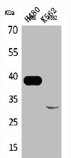
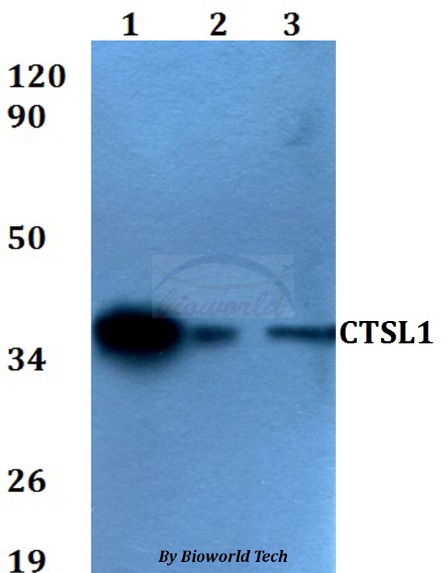
Western Blot analysis of H460 K562 cells using Cathepsin L Polyclonal Antibody
Western Blot analysis of H460 K562 cells using Cathepsin L Polyclonal Antibody
CTSL Antibody
CSB-PA004835
ApplicationsWestern Blot, ELISA
Product group Antibodies
ReactivityHuman
TargetCTSL
Overview
- SupplierCusabio
- Product NameCTSL Antibody
- Delivery Days Customer20
- ApplicationsWestern Blot, ELISA
- CertificationResearch Use Only
- ClonalityPolyclonal
- ConjugateUnconjugated
- Gene ID1514
- Target nameCTSL
- Target descriptioncathepsin L
- Target synonymsCATL, CTSL1, MEP, procathepsin L, cathepsin L1, major excreted protein
- HostRabbit
- IsotypeIgG
- Protein IDP07711
- Protein NameProcathepsin L
- ReactivityHuman
- Storage Instruction-20°C or -80°C
- UNSPSC41116161
Related products
Product group Antibodies
Anti-CTSL1 AntibodyA28775
ApplicationsWestern Blot
ReactivityHuman, Mouse, Rat
- SizePrice

Product group Antibodies
Anti-Cathepsin L/MEP/CTSL Antibody Picoband(r)A01589-2-CARRIER-FREE
ApplicationsWestern Blot
ReactivityHuman, Mouse
TargetCTSL
- SizePrice
Product group Antibodies
Cathepsin LVKH (9C3) Monoclonal AntibodyBSM-52919R
ApplicationsFlow Cytometry, Western Blot, ImmunoHistoChemistry, ImmunoHistoChemistry Paraffin
ReactivityHuman, Mouse, Rat
TargetCTSL
- SizePrice
Product group Antibodies
Ctsl Polyclonal AntibodyCAC11818
ApplicationsWestern Blot, ELISA
ReactivityMonkey
- SizePrice
Product group Antibodies
References
Cathepsin L antibody [33/2]GTX26314
ApplicationsWestern Blot, ELISA, ImmunoHistoChemistry, ImmunoHistoChemistry Frozen
ReactivityHuman, Mink, Mouse, Rat
TargetCTSL
- SizePrice
Product group Antibodies
Anti-CTSL AntibodyHPA070413
ApplicationsWestern Blot, ImmunoCytoChemistry
ReactivityHuman
TargetCTSL
- SizePrice
Product group Antibodies

CTSL / Cathepsin L AntibodyLS-C400630
ApplicationsELISA, ImmunoHistoChemistry
ReactivityHuman
TargetCTSL
- SizePrice