

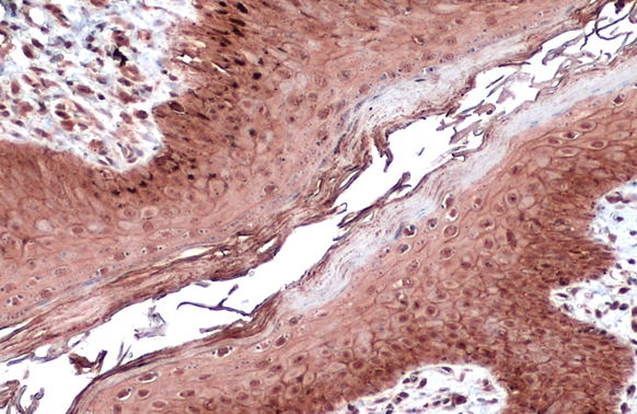
E-Cadherin antibody detects E-Cadherin protein at cell membrane and cytoplasm by immunohistochemical analysis. Sample: Paraffin-embedded dog skin. E-Cadherin stained by E-Cadherin antibody (GTX124178) diluted at 1:500. Antigen Retrieval: Citrate buffer, pH 6.0, 15 min
 diluted at 1:1000.")
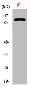
![Whole cell extracts (30 μg) was separated by 7.5% SDS-PAGE, and the membrane was blotted with E-Cadherin antibody [N3C2], Internal (GTX124178) diluted at 1:5000. The HRP-conjugated anti-rabbit IgG antibody (GTX213110-01) was used to detect the primary antibody.](https://www.genetex.com/upload/website/prouct_img/normal/GTX124178/GTX124178_43362_20190920_WB_D_22111423_152.webp "Whole cell extracts (30 μg) was separated by 7.5% SDS-PAGE, and the membrane was blotted with E-Cadherin antibody [N3C2], Internal (GTX124178) diluted at 1:5000. The HRP-conjugated anti-rabbit IgG antibody (GTX213110-01) was used to detect the primary antibody.")
![Various whole cell extracts (30 μg) were separated by 5% SDS-PAGE, and the membrane was blotted with E-Cadherin antibody [N3C2], Internal (GTX124178) diluted at 1:3000. The HRP-conjugated anti-rabbit IgG antibody (GTX213110-01) was used to detect the primary antibody. Corresponding RNA expression data for the same cell lines are based on Human Protein Atlas program.](https://www.genetex.com/upload/website/prouct_img/normal/GTX124178/GTX124178_43362_20191025_WB_TPM_watermark_w_23060522_216.webp "Various whole cell extracts (30 μg) were separated by 5% SDS-PAGE, and the membrane was blotted with E-Cadherin antibody [N3C2], Internal (GTX124178) diluted at 1:3000. The HRP-conjugated anti-rabbit IgG antibody (GTX213110-01) was used to detect the primary antibody. Corresponding RNA expression data for the same cell lines are based on Human Protein Atlas program.")
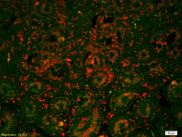
![E-Cadherin antibody [N3C2], Internal detects E-Cadherin protein at cell membrane by immunofluorescent analysis. Sample: HCT116 cells were fixed in 4% paraformaldehyde at RT for 15 min. Green: E-Cadherin stained by E-Cadherin antibody [N3C2], Internal (GTX124178) diluted at 1:500. Blue: Fluoroshield with DAPI (GTX30920). Scale bar= 10 μm.](https://www.genetex.com/upload/website/prouct_img/normal/GTX124178/GTX124178_43754_20200806_ICC_IF_w_23060522_989.webp "E-Cadherin antibody [N3C2], Internal detects E-Cadherin protein at cell membrane by immunofluorescent analysis. Sample: HCT116 cells were fixed in 4% paraformaldehyde at RT for 15 min. Green: E-Cadherin stained by E-Cadherin antibody [N3C2], Internal (GTX124178) diluted at 1:500. Blue: Fluoroshield with DAPI (GTX30920). Scale bar= 10 μm.")
![Immunoprecipitation of E-Cadherin protein from MCF-7 whole cell extracts using 5 μg of E-Cadherin antibody [N3C2], Internal (GTX124178). Western blot analysis was performed using E-Cadherin antibody [N3C2], Internal (GTX124178). EasyBlot anti-Rabbit IgG (GTX221666-01) was used as a secondary reagent.](https://www.genetex.com/upload/website/prouct_img/normal/GTX124178/GTX124178_40835_20151016_IP_w_23060522_351.webp "Immunoprecipitation of E-Cadherin protein from MCF-7 whole cell extracts using 5 μg of E-Cadherin antibody [N3C2], Internal (GTX124178). Western blot analysis was performed using E-Cadherin antibody [N3C2], Internal (GTX124178). EasyBlot anti-Rabbit IgG (GTX221666-01) was used as a secondary reagent.")
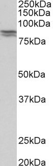
![E-Cadherin antibody [N3C2], Internal detects E-Cadherin protein by western blot analysis. Whole cell extracts (30 μg) was separated by 7.5% SDS-PAGE, and the membrane was blotted with E-Cadherin antibody [N3C2], Internal (GTX124178) diluted at 1:3000. The HRP-conjugated anti-rabbit IgG antibody (GTX213110-01) was used to detect the primary antibody.](https://www.genetex.com/upload/website/prouct_img/normal/GTX124178/GTX124178_40835_20151015_WB_w_23060522_185.webp "E-Cadherin antibody [N3C2], Internal detects E-Cadherin protein by western blot analysis. Whole cell extracts (30 μg) was separated by 7.5% SDS-PAGE, and the membrane was blotted with E-Cadherin antibody [N3C2], Internal (GTX124178) diluted at 1:3000. The HRP-conjugated anti-rabbit IgG antibody (GTX213110-01) was used to detect the primary antibody.")
![Various whole cell extracts (30 μg) were separated by 5% SDS-PAGE, and the membranes were blotted with E-Cadherin antibody [N3C2], Internal (GTX124178) diluted at 1:3000 and competitor's antibody diluted at 1:3000. The HRP-conjugated anti-rabbit IgG antibody (GTX213110-01) was used to detect the primary antibody. *The competitor is not affiliated with GeneTex and does not endorse this product.](https://www.genetex.com/upload/website/prouct_img/normal/GTX124178/GTX124178_40835_20200131_WB_competitor_watermark_w_23060522_322.webp "Various whole cell extracts (30 μg) were separated by 5% SDS-PAGE, and the membranes were blotted with E-Cadherin antibody [N3C2], Internal (GTX124178) diluted at 1:3000 and competitor's antibody diluted at 1:3000. The HRP-conjugated anti-rabbit IgG antibody (GTX213110-01) was used to detect the primary antibody. *The competitor is not affiliated with GeneTex and does not endorse this product.")
![Non-transfected (–) and transfected (+) MCF-7 whole cell extracts (30 μg) were separated by 5% SDS-PAGE, and the membrane was blotted with E-Cadherin antibody [N3C2], Internal (GTX124178) diluted at 1:10000. The HRP-conjugated anti-rabbit IgG antibody (GTX213110-01) was used to detect the primary antibody.](https://www.genetex.com/upload/website/prouct_img/normal/GTX124178/GTX124178_40835_20160602_WB_shRNA_watermark_w_23060522_997.webp "Non-transfected (–) and transfected (+) MCF-7 whole cell extracts (30 μg) were separated by 5% SDS-PAGE, and the membrane was blotted with E-Cadherin antibody [N3C2], Internal (GTX124178) diluted at 1:10000. The HRP-conjugated anti-rabbit IgG antibody (GTX213110-01) was used to detect the primary antibody.")
![Various whole cell extracts (30 μg) were separated by 7.5% SDS-PAGE, and the membrane was blotted with E-Cadherin antibody [N3C2], Internal (GTX124178) diluted at 1:3000. The HRP-conjugated anti-rabbit IgG antibody (GTX213110-01) was used to detect the primary antibody. Corresponding RNA expression data for the same cell lines are based on Human Protein Atlas program.](https://www.genetex.com/upload/website/prouct_img/normal/GTX124178/GTX124178_45175_20230922_WB_TPM_watermark_24082301_417.webp "Various whole cell extracts (30 μg) were separated by 7.5% SDS-PAGE, and the membrane was blotted with E-Cadherin antibody [N3C2], Internal (GTX124178) diluted at 1:3000. The HRP-conjugated anti-rabbit IgG antibody (GTX213110-01) was used to detect the primary antibody. Corresponding RNA expression data for the same cell lines are based on Human Protein Atlas program.")
E-Cadherin antibody detects E-Cadherin protein at cell membrane and cytoplasm by immunohistochemical analysis. Sample: Paraffin-embedded dog skin. E-Cadherin stained by E-Cadherin antibody (GTX124178) diluted at 1:500. Antigen Retrieval: Citrate buffer, pH 6.0, 15 min
E-Cadherin antibody [N3C2], Internal
GTX124178
ApplicationsImmunoFluorescence, ImmunoPrecipitation, Western Blot, ImmunoCytoChemistry, ImmunoHistoChemistry, ImmunoHistoChemistry Paraffin
Product group Antibodies
ReactivityCanine, Human, Rat
TargetCDH1
Overview
- SupplierGeneTex
- Product NameE-Cadherin antibody [N3C2], Internal
- Delivery Days Customer9
- Application Supplier NoteWB: 1:500-1:10000. ICC/IF: 1:100-1:1000. IP: 1:100-1:500. *Optimal dilutions/concentrations should be determined by the researcher.Not tested in other applications.
- ApplicationsImmunoFluorescence, ImmunoPrecipitation, Western Blot, ImmunoCytoChemistry, ImmunoHistoChemistry, ImmunoHistoChemistry Paraffin
- CertificationResearch Use Only
- ClonalityPolyclonal
- Concentration1.09 mg/ml
- ConjugateUnconjugated
- Gene ID999
- Target nameCDH1
- Target descriptioncadherin 1
- Target synonymsArc-1, BCDS1, CD324, CDHE, ECAD, LCAM, UVO, cadherin-1, CAM 120/80, E-cadherin 1, cadherin 1, E-cadherin (epithelial), cadherin 1, type 1, E-cadherin (epithelial), calcium-dependent adhesion protein, epithelial, cell-CAM 120/80, epididymis secretory sperm binding protein, epithelial cadherin, uvomorulin
- HostRabbit
- IsotypeIgG
- Protein IDP12830
- Protein NameCadherin-1
- Scientific DescriptionThis gene is a classical cadherin from the cadherin superfamily. The encoded protein is a calcium dependent cell-cell adhesion glycoprotein comprised of five extracellular cadherin repeats, a transmembrane region and a highly conserved cytoplasmic tail. Mutations in this gene are correlated with gastric, breast, colorectal, thyroid and ovarian cancer. Loss of function is thought to contribute to progression in cancer by increasing proliferation, invasion, and/or metastasis. The ectodomain of this protein mediates bacterial adhesion to mammalian cells and the cytoplasmic domain is required for internalization. Identified transcript variants arise from mutation at consensus splice sites. [provided by RefSeq]
- ReactivityCanine, Human, Rat
- Storage Instruction-20°C or -80°C,2°C to 8°C
- UNSPSC41116161
Datasheet
Related products
Product group Antibodies
Anti-E Cadherin AntibodyA83532
ApplicationsWestern Blot, ELISA, ImmunoHistoChemistry
ReactivityHuman, Mouse, Porcine, Rat
- SizePrice
Product group Antibodies
Anti-CDH1 Antibody144-03044
ApplicationsImmunoFluorescence, Western Blot, ImmunoHistoChemistry
ReactivityHuman, Mouse, Rat
TargetCDH1
- SizePrice
Product group Antibodies

Anti-E Cadherin [4A2]AB01700-1.1-BT
ApplicationsFlow Cytometry, ImmunoFluorescence, ImmunoPrecipitation, Western Blot, ImmunoHistoChemistry
ReactivityHuman, Mouse, Rat
TargetCDH1
- SizePrice

Product group Antibodies
Anti-CDH1 AntibodyAMAB90862
ApplicationsWestern Blot, ImmunoCytoChemistry, ImmunoHistoChemistry
ReactivityHuman
TargetCDH1
- SizePrice
Product group Antibodies
References
E cadherin Polyclonal AntibodyBS-10009R
ApplicationsFlow Cytometry, ImmunoFluorescence, Western Blot, ELISA, ImmunoCytoChemistry, ImmunoHistoChemistry, ImmunoHistoChemistry Frozen, ImmunoHistoChemistry Paraffin
ReactivityBovine, Equine, Human, Mouse, Porcine, Rabbit, Rat
TargetCDH1
- SizePrice
Product group Antibodies
CDH1 AntibodyCSB-PA002243
ApplicationsImmunoFluorescence, Western Blot, ELISA, ImmunoHistoChemistry
ReactivityHuman
TargetCDH1
- SizePrice
Product group Antibodies

Goat anti-CDH1 (aa662-675)EB11206
ApplicationsWestern Blot, ELISA, ImmunoHistoChemistry
ReactivityCanine, Human, Mouse, Porcine, Rat
TargetCDH1
- SizePrice
Product group Antibodies
Cdh1 Polyclonal AntibodyCAC11436
ApplicationsImmunoFluorescence, ImmunoPrecipitation, Western Blot, ELISA, ImmunoHistoChemistry
TargetCDH1
- SizePrice
Product group Antibodies

ApplicationsFlow Cytometry, Western Blot, ImmunoCytoChemistry, ImmunoHistoChemistry, ImmunoHistoChemistry Frozen
ReactivityHuman
TargetCDH1
- SizePrice