

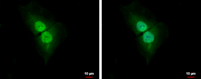
Cyclin D1 antibody [N2C3] detects Cyclin D1 protein at cytoplasm and nucleus by immunofluorescent analysis. Sample: SKNSH cells were fixed in 4% paraformaldehyde at RT for 15 min. Green: Cyclin D1 protein stained by Cyclin D1 antibody [N2C3] (GTX112874) diluted at 1:500. Blue: Hoechst 33342 staining.
![Cyclin D1 antibody [N2C3] detects Cyclin D1 protein at nucleus by immunohistochemical analysis. Sample: Paraffin-embedded human breast carcinoma. Cyclin D1 stained by Cyclin D1 antibody [N2C3] (GTX112874) diluted at 1:500. Antigen Retrieval: Citrate buffer, pH 6.0, 15 min](https://www.genetex.com/upload/website/prouct_img/normal/GTX112874/GTX112874_41073_20180928_IHC-P_w_23060500_705.webp "Cyclin D1 antibody [N2C3] detects Cyclin D1 protein at nucleus by immunohistochemical analysis. Sample: Paraffin-embedded human breast carcinoma. Cyclin D1 stained by Cyclin D1 antibody [N2C3] (GTX112874) diluted at 1:500. Antigen Retrieval: Citrate buffer, pH 6.0, 15 min")


Cyclin D1 antibody [N2C3] detects Cyclin D1 protein at cytoplasm and nucleus by immunofluorescent analysis. Sample: SKNSH cells were fixed in 4% paraformaldehyde at RT for 15 min. Green: Cyclin D1 protein stained by Cyclin D1 antibody [N2C3] (GTX112874) diluted at 1:500. Blue: Hoechst 33342 staining.
Cyclin D1 antibody [N2C3]
GTX112874
ApplicationsImmunoFluorescence, Western Blot, ImmunoCytoChemistry, ImmunoHistoChemistry, ImmunoHistoChemistry Frozen, ImmunoHistoChemistry Paraffin
Product group Antibodies
ReactivityHuman, Mouse
TargetCCND1
Overview
- SupplierGeneTex
- Product NameCyclin D1 antibody [N2C3]
- Delivery Days Customer9
- Application Supplier NoteICC/IF: 1:100-1:1000. IHC-P: 1:100-1:1000. *Optimal dilutions/concentrations should be determined by the researcher.Not tested in other applications.
- ApplicationsImmunoFluorescence, Western Blot, ImmunoCytoChemistry, ImmunoHistoChemistry, ImmunoHistoChemistry Frozen, ImmunoHistoChemistry Paraffin
- CertificationResearch Use Only
- ClonalityPolyclonal
- Concentration1 mg/ml
- ConjugateUnconjugated
- Gene ID595
- Target nameCCND1
- Target descriptioncyclin D1
- Target synonymsBCL1, D11S287E, PRAD1, U21B31, G1/S-specific cyclin-D1, B-cell CLL/lymphoma 1, B-cell lymphoma 1 protein, BCL-1 oncogene, PRAD1 oncogene
- HostRabbit
- IsotypeIgG
- Protein IDP24385
- Protein NameG1/S-specific cyclin-D1
- Scientific DescriptionThe protein encoded by this gene belongs to the highly conserved cyclin family, whose members are characterized by a dramatic periodicity in protein abundance throughout the cell cycle. Cyclins function as regulators of CDK kinases. Different cyclins exhibit distinct expression and degradation patterns which contribute to the temporal coordination of each mitotic event. This cyclin forms a complex with and functions as a regulatory subunit of CDK4 or CDK6, whose activity is required for cell cycle G1/S transition. This protein has been shown to interact with tumor suppressor protein Rb and the expression of this gene is regulated positively by Rb. Mutations, amplification and overexpression of this gene, which alters cell cycle progression, are observed frequently in a variety of tumors and may contribute to tumorigenesis. [provided by RefSeq]
- ReactivityHuman, Mouse
- Storage Instruction-20°C or -80°C,2°C to 8°C
- UNSPSC12352203
References
- Yeh HT, Tsai YS, Chen MS, et al. Flavopereirine induces cell cycle arrest and apoptosis via the AKT/p38 MAPK/ERK1/2 signaling pathway in human breast cancer cells. Eur J Pharmacol. 2019,863:172658. doi: 10.1016/j.ejphar.2019.172658Read this paper
- Chang YC, Fong Y, Tsai EM, et al. Exogenous C₈-Ceramide Induces Apoptosis by Overproduction of ROS and the Switch of Superoxide Dismutases SOD1 to SOD2 in Human Lung Cancer Cells. Int J Mol Sci. 2018,19(10). doi: 10.3390/ijms19103010Read this paper
- Liang ML, Hsieh TH, Liu YR, et al. Significance of cyclin D1 overexpression in progression and radio-resistance of pediatric ependymomas. Oncotarget. 2018,9(2):2527-2542. doi: 10.18632/oncotarget.23509Read this paper
- Yuan SY, Shiau MY, Ou YC, et al. Miconazole induces apoptosis via the death receptor 5-dependent and mitochondrial-mediated pathways in human bladder cancer cells. Oncol Rep. 2017,37(6):3606-3616. doi: 10.3892/or.2017.5608Read this paper
- Chang WT, Fong Y, Chuang SC, et al. 9-bis[2-(pyrrolidin-1-yl)ethoxy]-6-{4-[2-(pyrrolidin-1-yl)ethoxy]phenyl}-11H-indeno[1, 2-c]quinolin-11-one (BPIQ), A Quinoline Derivative Inhibits Human Hepatocellular Carcinoma Cells by Inducing ER Stress and Apoptosis. Anticancer Agents Med Chem. 2017,17(5):692-700. doi: 10.2174/1871520616666160802121456Read this paper
- Lee WJ, Jo SY, Lee MH, et al. The Effect of MCP-1/CCR2 on the Proliferation and Senescence of Epidermal Constituent Cells in Solar Lentigo. Int J Mol Sci. 2016,17(6). doi: 10.3390/ijms17060948Read this paper
- Luo CW, Wu CC, Ch'ang HJ. Radiation sensitization of tumor cells induced by shear stress: the roles of integrins and FAK. Biochim Biophys Acta. 2014,1843(9):2129-37. doi: 10.1016/j.bbamcr.2014.06.007Read this paper
- Lin CY, Tsai PH, Kandaswami CC, et al. Role of tissue transglutaminase 2 in the acquisition of a mesenchymal-like phenotype in highly invasive A431 tumor cells. Mol Cancer. 2011,10:87. doi: 10.1186/1476-4598-10-87Read this paper
- Pan J, Nakade K, Huang YC, et al. Suppression of cell-cycle progression by Jun dimerization protein-2 (JDP2) involves downregulation of cyclin-A2. Oncogene. 2010,29(47):6245-56. doi: 10.1038/onc.2010.355Read this paper
Datasheet
Related products
Product group Antibodies

CCND1-Y226 AntibodyABX032474
ApplicationsImmunoFluorescence, Western Blot, ELISA, ImmunoCytoChemistry
- SizePrice
Product group Antibodies
Anti-CCND1 Antibody144-11022
ApplicationsImmunoFluorescence, ImmunoPrecipitation, Western Blot, ImmunoHistoChemistry
ReactivityHuman, Mouse, Rat
TargetCCND1
- SizePrice

Product group Antibodies
Anti-CCND1 AntibodyAMAB91779
ApplicationsImmunoCytoChemistry
ReactivityHuman
TargetCCND1
- SizePrice
![IHC-P analysis of human breast tissue using GTX04738 Cyclin D1 antibody [SP4]. Antigen retrieval : EDTA Buffer pH 8.0_x000D_
_x000D_](https://www.genetex.com/upload/website/prouct_img/normal/GTX04738/GTX04738_20240325_IHC-P_24032422_761.webp)
Product group Antibodies
Cyclin D1 antibody [SP4]GTX04738
ApplicationsImmunoHistoChemistry, ImmunoHistoChemistry Paraffin
ReactivityHuman
TargetCCND1
- SizePrice
Product group Antibodies
References
Cyclin D1 antibody [N1C3]GTX108624
ApplicationsImmunoFluorescence, ImmunoPrecipitation, Western Blot, ELISA, ImmunoCytoChemistry, ImmunoHistoChemistry, ImmunoHistoChemistry Paraffin
ReactivityCanine, Feline, Human, Rat
TargetCCND1
- SizePrice
![Wild-type (WT) and Cyclin D1 knockout (KO) HeLa cell extracts (30 μg) were separated by 12% SDS-PAGE, and the membrane was blotted with Cyclin D1 antibody [N1C3-2] (GTX110541) diluted at 1:1000. The HRP-conjugated anti-rabbit IgG antibody (GTX213110-01) was used to detect the primary antibody, and the signal was developed with Trident ECL plus-Enhanced.](https://www.genetex.com/upload/website/prouct_img/normal/GTX110541/GTX110541_40450_20170504_KO_watermark_w_23060500_976.webp)
Product group Antibodies
Cyclin D1 antibody [N1C3-2]GTX110541
ApplicationsWestern Blot
ReactivityHuman
TargetCCND1
- SizePrice
Product group Antibodies
References
Cyclin D1 antibody [GT8912]GTX634347
ApplicationsWestern Blot, ELISA
ReactivityHuman
TargetCCND1
- SizePrice
![Wild-type (WT) and CCND1 knockout (KO) HeLa cell extracts (30 μg) were separated by 12% SDS-PAGE, and the membrane was blotted with Cyclin D1 antibody [HL2219] (GTX638223) diluted at 1:1000. The HRP-conjugated anti-rabbit IgG antibody (GTX213110-01) was used to detect the primary antibody.](https://www.genetex.com/upload/website/prouct_img/normal/GTX638223/GTX638223_T-44942_20230303_WB_KO_watermark_23030717_165.webp)
Product group Antibodies
Cyclin D1 antibody [HL2219]GTX638223
ApplicationsWestern Blot
ReactivityCanine, Human, Rat
TargetCCND1
- SizePrice
Product group Antibodies
ApplicationsWestern Blot
ReactivityMouse, Rat
TargetCCND1
- SizePrice