

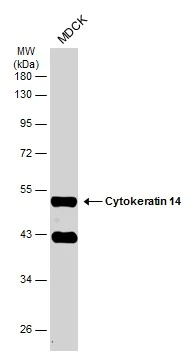
Whole cell extract (30 μg) was separated by 10% SDS-PAGE, and the membrane was blotted with Cytokeratin 14 antibody (GTX104124) diluted at 1:6000. The HRP-conjugated anti-rabbit IgG antibody (GTX213110-01) was used to detect the primary antibody.
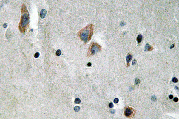
 antibody at 1:100 dilution.
Antigen Retrieval: Trilogy? (EDTA based, pH 8.0) buffer, 15min")
![Cytokeratin 14 antibody detects Cytokeratin 14 protein at cytoplasm by immunofluorescent analysis. Sample: HeLa cells were fixed in 4% paraformaldehyde at RT for 15 min. Green: Cytokeratin 14 protein stained by Cytokeratin 14 antibody (GTX104124) diluted at 1:200. Red: alpha Tubulin, a cytoskeleton marker, stained by alpha Tubulin antibody [GT114] (GTX628802) diluted at 1:1000. Blue: Hoechst 33342 staining.](https://www.genetex.com/upload/website/prouct_img/normal/GTX104124/GTX104124_39848_20150410_IFA_w_23060120_451.webp "Cytokeratin 14 antibody detects Cytokeratin 14 protein at cytoplasm by immunofluorescent analysis. Sample: HeLa cells were fixed in 4% paraformaldehyde at RT for 15 min. Green: Cytokeratin 14 protein stained by Cytokeratin 14 antibody (GTX104124) diluted at 1:200. Red: alpha Tubulin, a cytoskeleton marker, stained by alpha Tubulin antibody [GT114] (GTX628802) diluted at 1:1000. Blue: Hoechst 33342 staining.")


 was separated by 10% SDS-PAGE, and the membrane was blotted with Cytokeratin 14 antibody (GTX104124) diluted at 1:6000.")
Whole cell extract (30 μg) was separated by 10% SDS-PAGE, and the membrane was blotted with Cytokeratin 14 antibody (GTX104124) diluted at 1:6000. The HRP-conjugated anti-rabbit IgG antibody (GTX213110-01) was used to detect the primary antibody.
Cytokeratin 14 antibody
GTX104124
ApplicationsImmunoFluorescence, Western Blot, ImmunoCytoChemistry, ImmunoHistoChemistry, ImmunoHistoChemistry Frozen, ImmunoHistoChemistry Paraffin
Product group Antibodies
ReactivityCanine, Human, Rat
TargetKRT14
Overview
- SupplierGeneTex
- Product NameCytokeratin 14 antibody
- Delivery Days Customer9
- Application Supplier NoteWB: 1:1000-1:10000. ICC/IF: 1:100-1:1000. IHC-P: 1:100-1:1000. *Optimal dilutions/concentrations should be determined by the researcher.Not tested in other applications.
- ApplicationsImmunoFluorescence, Western Blot, ImmunoCytoChemistry, ImmunoHistoChemistry, ImmunoHistoChemistry Frozen, ImmunoHistoChemistry Paraffin
- CertificationResearch Use Only
- ClonalityPolyclonal
- Concentration0.51 mg/ml
- ConjugateUnconjugated
- Gene ID3861
- Target nameKRT14
- Target descriptionkeratin 14
- Target synonymsCK14, EBS1, EBS1A, EBS1B, EBS1C, EBS1D, EBS3, EBS4, K14, NFJ, keratin, type I cytoskeletal 14, cytokeratin 14, keratin 14, type I
- HostRabbit
- IsotypeIgG
- Protein IDP02533
- Protein NameKeratin, type I cytoskeletal 14
- Scientific DescriptionThis gene encodes a member of the keratin family, the most diverse group of intermediate filaments. This gene product, a type I keratin, is usually found as a heterotetramer with two keratin 5 molecules, a type II keratin. Together they form the cytoskeleton of epithelial cells. Mutations in the genes for these keratins are associated with epidermolysis bullosa simplex. At least one pseudogene has been identified at 17p12-p11. [provided by RefSeq]
- ReactivityCanine, Human, Rat
- Storage Instruction-20°C or -80°C,2°C to 8°C
- UNSPSC41116161
Datasheet
Related products
Product group Antibodies
ApplicationsELISA, ImmunoHistoChemistry
ReactivityHuman, Mouse, Rat
- SizePrice
Product group Antibodies

Anti-Cytokeratin 14 [RCK107]AB03338-1.1
ApplicationsFlow Cytometry, Western Blot, ImmunoHistoChemistry
ReactivityCanine, Human, Porcine, Rat
TargetKRT14
- SizePrice
Product group Antibodies
Anti-Cytokeratin 14 Antibody118-10011
ApplicationsELISA, ImmunoHistoChemistry
ReactivityHuman
- SizePrice
Product group Antibodies
Anti-KRT14 AntibodyAMAB91968
ApplicationsWestern Blot, ImmunoCytoChemistry, ImmunoHistoChemistry
ReactivityHuman
TargetKRT14
- SizePrice

Product group Antibodies
Anti-Cytokeratin 14/KRT14 Antibody Picoband(r)A01432-CARRIER-FREE
ApplicationsFlow Cytometry, Western Blot, ImmunoCytoChemistry, ImmunoHistoChemistry, ImmunoHistoChemistry Frozen
ReactivityHuman, Mouse, Rat
TargetKRT14
- SizePrice
Product group Antibodies
Cytokeratin 13 Recombinant AntibodyBSM-52053R
ApplicationsImmunoFluorescence, Western Blot, ImmunoCytoChemistry, ImmunoHistoChemistry, ImmunoHistoChemistry Frozen, ImmunoHistoChemistry Paraffin
ReactivityHuman, Mouse
TargetKRT14
- SizePrice
Product group Antibodies

Cytokeratin 14CK514
ApplicationsImmunoHistoChemistry, ImmunoHistoChemistry Frozen, ImmunoHistoChemistry Paraffin
ReactivityHuman
TargetKRT14
- SizePrice
Product group Antibodies
ApplicationsImmunoPrecipitation, Western Blot, ImmunoCytoChemistry, ImmunoHistoChemistry
ReactivityRat
TargetKRT14
- SizePrice
Product group Antibodies
KRT14 AntibodyCSB-PA005038
ApplicationsWestern Blot, ELISA
ReactivityHuman, Mouse, Rat
TargetKRT14
- SizePrice