

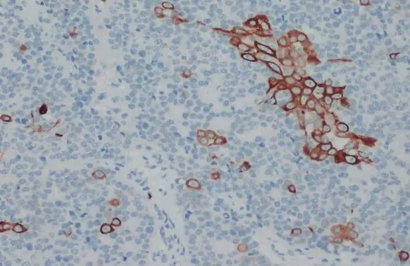
Cytokeratin 18 antibody detects Cytokeratin 18 protein at cytoskeleton by immunohistochemical analysis. Sample: Paraffin-embedded dog malignant carcinoma. Cytokeratin 18 stained by Cytokeratin 18 antibody (GTX105624) diluted at 1:500. Antigen Retrieval: Citrate buffer, pH 6.0, 15 min
 diluted at 1:500. Antigen Retrieval: Citrate buffer, pH 6.0, 15 min")
 diluted at 1:500.")
 diluted at 1:500. Antigen Retrieval: Citrate buffer, pH 6.0, 15 min")
 diluted at 1:500. Antigen Retrieval: Citrate buffer, pH 6.0, 15 min")
 diluted at 1:500. Antigen Retrieval: Citrate buffer, pH 6.0, 15 min")
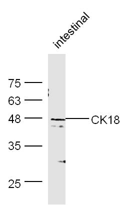
 was separated by 10% SDS-PAGE, and the membrane was blotted with Cytokeratin 18 antibody (GTX105624) diluted at 1:2000. The HRP-conjugated anti-rabbit IgG antibody (GTX213110-01) was used to detect the primary antibody, and the signal was developed with Trident ECL plus-Enhanced.")
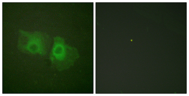
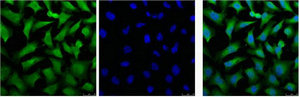
![Cytokeratin 18 antibody [N2C2], Internal detects Cytokeratin 18 protein at cytoskeleton by immunofluorescent analysis. Sample: HeLa cells were fixed in ice-cold MeOH for 5 min. Green: Cytokeratin 18 protein stained by Cytokeratin 18 antibody [N2C2], Internal (GTX105624) diluted at 1:200. Blue: Hoechst 33342 staining. Scale bar = 10 μm.](https://www.genetex.com/upload/website/prouct_img/normal/GTX105624/GTX105624_39890_IFA_w_23060120_323.webp "Cytokeratin 18 antibody [N2C2], Internal detects Cytokeratin 18 protein at cytoskeleton by immunofluorescent analysis. Sample: HeLa cells were fixed in ice-cold MeOH for 5 min. Green: Cytokeratin 18 protein stained by Cytokeratin 18 antibody [N2C2], Internal (GTX105624) diluted at 1:200. Blue: Hoechst 33342 staining. Scale bar = 10 μm.")
![Non-transfected (–) and transfected (+) HeLa whole cell extracts (15 μg) were separated by 10% SDS-PAGE, and the membrane was blotted with Cytokeratin 18 antibody [N2C2], Internal (GTX105624) diluted at 1:2000.](https://www.genetex.com/upload/website/prouct_img/normal/GTX105624/GTX105624_39890_20161006_WB_shRNA_watermark_w_23060120_237.webp "Non-transfected (–) and transfected (+) HeLa whole cell extracts (15 μg) were separated by 10% SDS-PAGE, and the membrane was blotted with Cytokeratin 18 antibody [N2C2], Internal (GTX105624) diluted at 1:2000.")
 A:H1299 B:Hep G2(GTX27900) 7.5% SDS PAGE GTX105624 diluted at 1:3000")
Cytokeratin 18 antibody detects Cytokeratin 18 protein at cytoskeleton by immunohistochemical analysis. Sample: Paraffin-embedded dog malignant carcinoma. Cytokeratin 18 stained by Cytokeratin 18 antibody (GTX105624) diluted at 1:500. Antigen Retrieval: Citrate buffer, pH 6.0, 15 min
Cytokeratin 18 antibody [N2C2], Internal
GTX105624
ApplicationsImmunoFluorescence, ImmunoPrecipitation, Western Blot, ImmunoCytoChemistry, ImmunoHistoChemistry, ImmunoHistoChemistry Paraffin
Product group Antibodies
ReactivityCanine, Human, Mouse
TargetKRT18
Overview
- SupplierGeneTex
- Product NameCytokeratin 18 antibody [N2C2], Internal
- Delivery Days Customer9
- Application Supplier NoteWB: 1:500-1:3000. ICC/IF: 1:100-1:1000. IHC-P: 1:100-1:1000. *Optimal dilutions/concentrations should be determined by the researcher.Not tested in other applications.
- ApplicationsImmunoFluorescence, ImmunoPrecipitation, Western Blot, ImmunoCytoChemistry, ImmunoHistoChemistry, ImmunoHistoChemistry Paraffin
- CertificationResearch Use Only
- ClonalityPolyclonal
- Concentration0.54 mg/ml
- ConjugateUnconjugated
- Gene ID3875
- Target nameKRT18
- Target descriptionkeratin 18
- Target synonymsCK-18, CYK18, K18, keratin, type I cytoskeletal 18, cell proliferation-inducing gene 46 protein, cytokeratin 18, keratin 18, type I
- HostRabbit
- IsotypeIgG
- Protein IDP05783
- Protein NameKeratin, type I cytoskeletal 18
- Scientific DescriptionKRT18 encodes the type I intermediate filament chain keratin 18. Keratin 18, together with its filament partner keratin 8, are perhaps the most commonly found members of the intermediate filament gene family. They are expressed in single layer epithelial tissues of the body. Mutations in this gene have been linked to cryptogenic cirrhosis. Two transcript variants encoding the same protein have been found for this gene. [provided by RefSeq]
- ReactivityCanine, Human, Mouse
- Storage Instruction-20°C or -80°C,2°C to 8°C
- UNSPSC41116161
Datasheet
Related products
Product group Antibodies
Anti-Keratin 18 AntibodyA93721
ApplicationsImmunoFluorescence, ImmunoPrecipitation, Western Blot, ELISA, ImmunoHistoChemistry
ReactivityHuman, Mouse, Rat
- SizePrice
Product group Antibodies

Anti-Cytokeratin 18 [RGE53]AB03339-1.1
ApplicationsFlow Cytometry, Western Blot, ImmunoHistoChemistry
ReactivityCanine, Chicken, Hamster, Human, Mouse, Porcine, Rabbit, Rat, Zebra Fish
TargetKRT18
- SizePrice

Product group Antibodies
Anti-Cytokeratin 18/KRT18 Antibody Picoband(r)A01357-1-CARRIER-FREE
ApplicationsFlow Cytometry, ImmunoFluorescence, Western Blot, ImmunoCytoChemistry, ImmunoHistoChemistry, ImmunoHistoChemistry Frozen
ReactivityHuman, Mouse, Rat
TargetKRT18
- SizePrice
Product group Antibodies
CK18 Polyclonal Antibodybs-1339R
ApplicationsFlow Cytometry, ImmunoFluorescence, Western Blot, ELISA, ImmunoCytoChemistry, ImmunoHistoChemistry, ImmunoHistoChemistry Frozen, ImmunoHistoChemistry Paraffin
ReactivityCanine, Chicken, Human, Mouse, Rabbit, Rat
TargetKRT18
- SizePrice
Product group Antibodies
KRT18 Monoclonal AntibodyCSB-MA000253
ApplicationsImmunoFluorescence, Western Blot, ELISA, ImmunoHistoChemistry
ReactivityHuman, Mouse, Rat
TargetKRT18
- SizePrice
Product group Antibodies

Goat anti-keratin 18EB12358
ApplicationsWestern Blot, ELISA, ImmunoHistoChemistry
ReactivityBovine, Canine, Human, Mouse, Porcine, Rat
TargetKRT18
- SizePrice
Product group Antibodies
ApplicationsFlow Cytometry
TargetKRT18
- SizePrice
![IHC-P analysis of human colon tissue using GTX20669 Cytokeratin 18 antibody [DA-7].](https://www.genetex.com/upload/website/prouct_img/normal/GTX20669/GTX20669_20191028_IHC-P_1_w_23060620_694.webp)
Product group Antibodies
Cytokeratin 18 antibody [DA-7]GTX20669
ApplicationsImmunoFluorescence, ImmunoPrecipitation, Western Blot, ELISA, ImmunoCytoChemistry, ImmunoHistoChemistry, ImmunoHistoChemistry Paraffin
ReactivityHuman
TargetKRT18
- SizePrice