

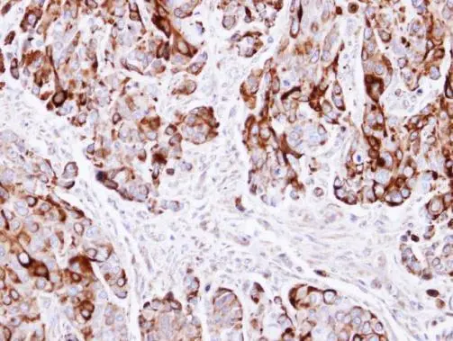
Immunohistochemical analysis of paraffin-embedded HT29 xenograft , using Cytokeratin 20(GTX100693) antibody at 1:500 dilution.
Antigen Retrieval: Trilogy? (EDTA based, pH 8.0) buffer, 15min
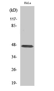
![Various whole cell extracts (30 μg) were separated by 10% SDS-PAGE, and the membrane was blotted with Cytokeratin 20 antibody [C2C3], C-term (GTX100693) diluted at 1:1000. The HRP-conjugated anti-rabbit IgG antibody (GTX213110-01) was used to detect the primary antibody. Corresponding RNA expression data for the same cell lines are based on Human Protein Atlas program.](https://www.genetex.com/upload/website/prouct_img/normal/GTX100693/GTX100693_39995_20231103_WB_TPM_watermark_23110819_786.webp "Various whole cell extracts (30 μg) were separated by 10% SDS-PAGE, and the membrane was blotted with Cytokeratin 20 antibody [C2C3], C-term (GTX100693) diluted at 1:1000. The HRP-conjugated anti-rabbit IgG antibody (GTX213110-01) was used to detect the primary antibody. Corresponding RNA expression data for the same cell lines are based on Human Protein Atlas program.")
Immunohistochemical analysis of paraffin-embedded HT29 xenograft , using Cytokeratin 20(GTX100693) antibody at 1:500 dilution.
Antigen Retrieval: Trilogy? (EDTA based, pH 8.0) buffer, 15min
Cytokeratin 20 antibody [C2C3], C-term
GTX100693
ApplicationsWestern Blot, ImmunoHistoChemistry, ImmunoHistoChemistry Paraffin
Product group Antibodies
ReactivityHuman
TargetKRT20
Overview
- SupplierGeneTex
- Product NameCytokeratin 20 antibody [C2C3], C-term
- Delivery Days Customer9
- Application Supplier NoteWB: 1:500-1:3000. IHC-P: 1:100-1:1000. *Optimal dilutions/concentrations should be determined by the researcher.Not tested in other applications.
- ApplicationsWestern Blot, ImmunoHistoChemistry, ImmunoHistoChemistry Paraffin
- CertificationResearch Use Only
- ClonalityPolyclonal
- Concentration1 mg/ml
- ConjugateUnconjugated
- Gene ID54474
- Target nameKRT20
- Target descriptionkeratin 20
- Target synonymsCD20, CK-20, CK20, K20, KRT21, keratin, type I cytoskeletal 20, cytokeratin 20, keratin 20, type I, protein IT
- HostRabbit
- IsotypeIgG
- Protein IDP35900
- Protein NameKeratin, type I cytoskeletal 20
- Scientific DescriptionThe protein encoded by this gene is a member of the keratin family. The keratins are intermediate filament proteins responsible for the structural integrity of epithelial cells and are subdivided into cytokeratins and hair keratins. The type I cytokeratins consist of acidic proteins which are arranged in pairs of heterotypic keratin chains. This cytokeratin is a major cellular protein of mature enterocytes and goblet cells and is specifically expressed in the gastric and intestinal mucosa. The type I cytokeratin genes are clustered in a region of chromosome 17q12-q21. [provided by RefSeq]
- ReactivityHuman
- Storage Instruction-20°C or -80°C,2°C to 8°C
- UNSPSC41116161
Datasheet
Related products
Product group Antibodies
ApplicationsImmunoFluorescence, Western Blot, ELISA, ImmunoHistoChemistry
ReactivityHuman, Mouse, Rat
- SizePrice
Product group Antibodies

Anti-Cytokeratin 20 [10F6B12A9]AB03189-1.1-BT
ApplicationsWestern Blot, ELISA, ImmunoHistoChemistry
ReactivityHuman
TargetKRT20
- SizePrice

Product group Antibodies
Anti-KRT20 AntibodyAMAB91564
ApplicationsImmunoHistoChemistry
ReactivityHuman
TargetKRT20
- SizePrice
Product group Antibodies
KRT20 Polyclonal AntibodyCAC15779
ApplicationsImmunoFluorescence, Western Blot, ELISA, ImmunoHistoChemistry
TargetKRT20
- SizePrice
Product group Antibodies
KRT20 AntibodyCSB-PA002048
ApplicationsImmunoFluorescence, Western Blot, ELISA, ImmunoHistoChemistry
ReactivityHuman, Mouse, Rat
TargetKRT20
- SizePrice
Product group Antibodies
ApplicationsImmunoFluorescence, Western Blot, ImmunoCytoChemistry, ImmunoHistoChemistry, ImmunoHistoChemistry Paraffin
ReactivityHuman, Mouse
TargetKRT20
- SizePrice
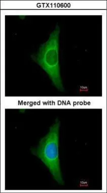
![ICC/IF analysis of A549 cells using DAPI (blue) and Cytokeratin 20 antibody [AT10D8] at a dilution of 1:200 (green).](https://www.genetex.com/upload/website/prouct_img/normal/GTX16500/GTX16500_ICCIF_w_23060620_604.webp)
Product group Antibodies
Cytokeratin 20 antibody [AT10D8]GTX16500
ApplicationsFlow Cytometry, ImmunoFluorescence, Western Blot, ELISA, ImmunoCytoChemistry
ReactivityHuman
TargetKRT20
- SizePrice
Product group Antibodies
ApplicationsWestern Blot, ImmunoHistoChemistry, ImmunoHistoChemistry Paraffin, Other Application
ReactivityHuman
TargetKRT20
- SizePrice