

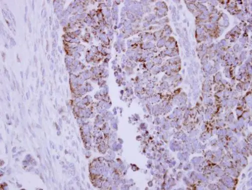
Immunohistochemical analysis of paraffin-embedded human ovarian carcinoma, using DLAT(GTX109766) antibody at 1:250 dilution.
Antigen Retrieval: Trilogy? (EDTA based, pH 8.0) buffer, 15min
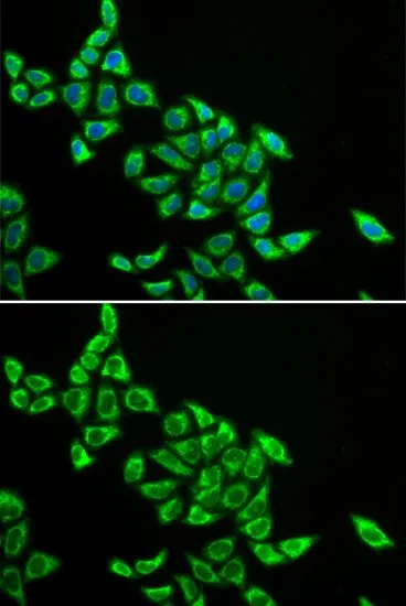
 diluted at 1:500. Blue: Hoechst 33342 staining.")
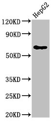
 were separated by 7.5% SDS-PAGE, and the membrane was blotted with DLAT antibody (GTX109766) diluted at 1:1000. The HRP-conjugated anti-rabbit IgG antibody (GTX213110-01) was used to detect the primary antibody.")
Immunohistochemical analysis of paraffin-embedded human ovarian carcinoma, using DLAT(GTX109766) antibody at 1:250 dilution.
Antigen Retrieval: Trilogy? (EDTA based, pH 8.0) buffer, 15min
DLAT antibody
GTX109766
ApplicationsImmunoFluorescence, Western Blot, ImmunoCytoChemistry, ImmunoHistoChemistry, ImmunoHistoChemistry Paraffin
Product group Antibodies
ReactivityHuman
TargetDLAT
Overview
- SupplierGeneTex
- Product NameDLAT antibody
- Delivery Days Customer9
- Application Supplier NoteWB: 1:5000-1:20000. ICC/IF: 1:100-1:1000. IHC-P: 1:100-1:1000. *Optimal dilutions/concentrations should be determined by the researcher.Not tested in other applications.
- ApplicationsImmunoFluorescence, Western Blot, ImmunoCytoChemistry, ImmunoHistoChemistry, ImmunoHistoChemistry Paraffin
- CertificationResearch Use Only
- ClonalityPolyclonal
- Concentration1 mg/ml
- ConjugateUnconjugated
- Gene ID1737
- Target nameDLAT
- Target descriptiondihydrolipoamide S-acetyltransferase
- Target synonymsDLTA, E2, PBC, PDC-E2, PDCE2, dihydrolipoyllysine-residue acetyltransferase component of pyruvate dehydrogenase complex, mitochondrial, 70 kDa mitochondrial autoantigen of primary biliary cirrhosis, E2 component of pyruvate dehydrogenase complex, M2 antigen complex 70 kDa subunit, dihydrolipoamide acetyltransferase component of pyruvate dehydrogenase complex, pyruvate dehydrogenase complex component E2
- HostRabbit
- IsotypeIgG
- Protein IDP10515
- Protein NameDihydrolipoyllysine-residue acetyltransferase component of pyruvate dehydrogenase complex, mitochondrial
- Scientific DescriptionThe DLAT gene encodes dihydrolipoamide acetyltransferase (EC 2.3.1.12), the E2 subunit of the mammalian pyruvate dehydrogenase complex (PDC; EC 1.2.4.1) of the inner mitochondrial membrane. Patients with primary biliary cirrhosis (PBC; MIM 109720) show autoantibodies to DLAT.[supplied by OMIM]
- ReactivityHuman
- Storage Instruction-20°C or -80°C,2°C to 8°C
- UNSPSC41116161
Datasheet
Related products
Product group Antibodies
DLAT AntibodyCSB-PA006926LA01HU
ApplicationsImmunoPrecipitation, Western Blot, ELISA
ReactivityHuman
TargetDLAT
- SizePrice
Product group Antibodies
Anti-DLAT Antibody144-60697
ApplicationsWestern Blot, ImmunoHistoChemistry
ReactivityHuman, Mouse, Rat
TargetDLAT
- SizePrice

Product group Antibodies
Anti-DLAT AntibodyA29546
ApplicationsWestern Blot, ImmunoHistoChemistry
ReactivityHuman, Mouse, Rat
- SizePrice
Product group Antibodies

DLAT / PDC-E2 AntibodyLS-C766087
ApplicationsWestern Blot, ELISA, ImmunoHistoChemistry
ReactivityHuman, Mouse, Rat
TargetDLAT
- SizePrice
Product group Antibodies
TargetDLAT
- SizePrice

Product group Antibodies
Anti-DLAT AntibodyHPA040786
ApplicationsWestern Blot, ImmunoCytoChemistry, ImmunoHistoChemistry
ReactivityHuman
TargetDLAT
- SizePrice

Product group Antibodies
Anti-DLAT Antibody Picoband(r)A04469-2-CARRIER-FREE
ApplicationsFlow Cytometry, ImmunoFluorescence, Western Blot, ELISA, ImmunoCytoChemistry, ImmunoHistoChemistry
ReactivityHuman, Mouse, Rat
TargetDLAT
- SizePrice
Product group Antibodies
DLAT antibody [4A4-B6-C10]GTX49202
ApplicationsImmunoFluorescence, ImmunoPrecipitation, Western Blot, ImmunoCytoChemistry
ReactivityHuman, Mouse
TargetDLAT
- SizePrice
Product group Antibodies
DLAT antibodyGTX54002
ApplicationsImmunoFluorescence, Western Blot, ImmunoCytoChemistry
ReactivityHuman, Mouse, Rat
TargetDLAT
- SizePrice