Bio-Connect
DOG-1 / TMEM16A (Gastrointestinal Stromal Tumor Marker)(DG1/2831R), CF647 conjugate, 0.1mg/mL [26628-22-8]

DOG-1 / TMEM16A (Gastrointestinal Stromal Tumor Marker)(DG1/2831R), CF647 conjugate, 0.1mg/mL [26628-22-8]

BNC472831
Biotium
ApplicationsImmunoHistoChemistry, ImmunoHistoChemistry Paraffin
Product group Antibodies
TargetANO1
Sign in to order and to see your custom pricing.
Large volume orders?
Order with a bulk request

Overview

  • Supplier
    Biotium
  • Product Name
    DOG-1 / TMEM16A (Gastrointestinal Stromal Tumor Marker)(DG1/2831R), CF647 conjugate, 0.1mg/mL
  • Delivery Days Customer
    9
  • Applications
    ImmunoHistoChemistry, ImmunoHistoChemistry Paraffin
  • Certification
    Research Use Only
  • Clonality
    Monoclonal
  • Clone ID
    DG1/2831R
  • Concentration
    0.1 mg/ml
  • Conjugate
    Other Conjugate
  • Gene ID55107
  • Target name
    ANO1
  • Target description
    anoctamin 1
  • Target synonyms
    anoctamin 1, calcium activated chloride channel; anoctamin-1; Ca2+-activated Cl- channel; calcium activated chloride channel; discovered on gastrointestinal stromal tumors protein 1; DOG1; oral cancer overexpressed 2; ORAOV2; TAOS2; TMEM16A; transmembrane protein 16A (eight membrane-spanning domains); tumor-amplified and overexpressed sequence 2
  • Host
    Rabbit
  • Isotype
    IgG
  • Scientific Description
    Expression of DOG-1 protein is elevated in the gastrointestinal stromal tumors (GISTs), c-kit signaling-driven mesenchymal tumors of the GI tract. DOG-1 is rarely expressed in other soft tissue tumors, which, due to appearance, may be difficult to diagnose. Immunoreactivity for DOG-1 has been reported in 97. 8 percent of scorable GISTs, including all c-kit negative GISTs. Overexpression of DOG-1 has been suggested to aid in the identification of GISTs, including Platelet-Derived Growth Factor Receptor Alpha mutants that fail to express c-kit antigen. The overall sensitivity of DOG1 and c-kit in GISTs is nearly identical: 94. 4% vs. 94. 7%. Primary antibodies are available purified, or with a selection of fluorescent CF® Dyes and other labels. CF® Dyes offer exceptional brightness and photostability. Note: Conjugates of blue fluorescent dyes like CF®405S and CF®405M are not recommended for detecting low abundance targets, because blue dyes have lower fluorescence and can give higher non-specific background than other dye colors.
  • Source
    Animal
  • Storage Instruction
    2°C to 8°C
  • UNSPSC
    12352203

Related products

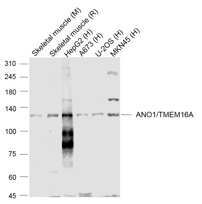
Lane 1: Mouse Skeletal muscle lysates; Lane 2: Rat Skeletal muscle lysates; Lane 3: Human HepG2 cell lysates; Lane 4: Human A673 cell lysates; Lane 5: Human U-2OS cell lysates; Lane 6: Human MKN45 cell lysates probed with TMEM16A Polyclonal Antibody, Unconjugated (bs-3794R) at 1:1000 dilution and 4˚C overnight incubation. Followed by conjugated secondary antibody incubation at 1:20000 for 60 min at 37˚C
Product group Antibodies
Bioss Antibodies
ApplicationsImmunoFluorescence, Western Blot, ELISA, ImmunoCytoChemistry, ImmunoHistoChemistry, ImmunoHistoChemistry Frozen, ImmunoHistoChemistry Paraffin
TargetANO1
  • SizePrice
Anti-ANO1 Antibody
Product group Antibodies
Antibodies.com
ApplicationsWestern Blot
  • SizePrice
Product group Antibodies
Absolute Antibody
Anti-DOG1 [19F2]Ab03308-10.0
ApplicationsWestern Blot, ELISA, ImmunoHistoChemistry
TargetANO1
  • SizePrice
Product group Antibodies
RayBiotech
ApplicationsImmunoFluorescence, Western Blot
TargetANO1
  • SizePrice
Product group Antibodies
MedChem Express
TargetANO1
  • SizePrice
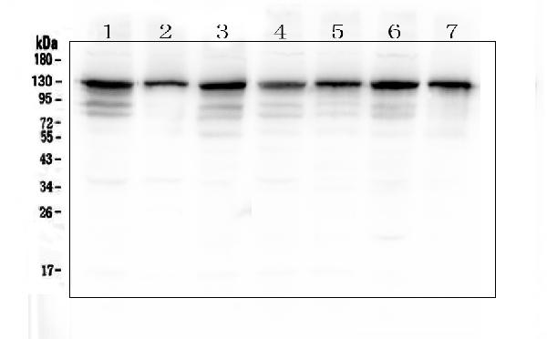
Figure 1. Western blot analysis of TMEM16A using anti-TMEM16A antibody (A00713). Electrophoresis was performed on a 5-20% SDS-PAGE gel at 70V (Stacking gel) / 90V (Resolving gel) for 2-3 hours. The sample well of each lane was loaded with 50ug of sample under reducing conditions. Lane 1: human Hela whole cell lysate, Lane 2: human HepG2 whole cell lysate, Lane 3: human A549 whole cell lysate, Lane 4: human PANC-1 whole cell lysate, Lane 5: human SK-OV-3 whole cell lysate, Lane 6: human SGC-7901 whole cell lysate, Lane 7: human COLO-320 whole cell lysate. After Electrophoresis, proteins were transferred to a Nitrocellulose membrane at 150mA for 50-90 minutes. Blocked the membrane with 5% Non-fat Milk/ TBS for 1.5 hour at RT. The membrane was incubated with rabbit anti-TMEM16A antigen affinity purified polyclonal antibody (Catalog # A00713) at 0.5 microg/mL overnight at 4°C, then washed with TBS-0.1%Tween 3 times with 5 minutes each and probed with a goat anti-rabbit IgG-HRP secondary antibody at a dilution of 1:10000 for 1.5 hour at RT. The signal is developed using an Enhanced Chemiluminescent detection (ECL) kit (Catalog # EK1002) with Tanon 5200 system. A specific band was detected for TMEM16A at approximately 130KD. The expected band size for TMEM16A is at 114KD.
Product group Antibodies
Boster Bio
ApplicationsFlow Cytometry, Western Blot, ImmunoHistoChemistry
TargetANO1
  • SizePrice
Product group Antibodies
Cusabio
ANO1 AntibodyCSB-PA030177
ApplicationsImmunoFluorescence, ELISA
ReactivityHuman
TargetANO1
  • SizePrice
Product group Antibodies
LifeSpan BioSciences
ApplicationsWestern Blot
TargetANO1
  • SizePrice
Immunohistochemistry analysis in human salivary gland and cerebral cortex tissues using HPA032148 antibody. Corresponding ANO1 RNA-seq data are presented for the same tissues.
Product group Antibodies
Atlas Antibodies
ApplicationsImmunoHistoChemistry
TargetANO1
  • SizePrice
Product group Antibodies
GeneTex
ApplicationsImmunoHistoChemistry, ImmunoHistoChemistry Paraffin
TargetANO1
  • SizePrice