

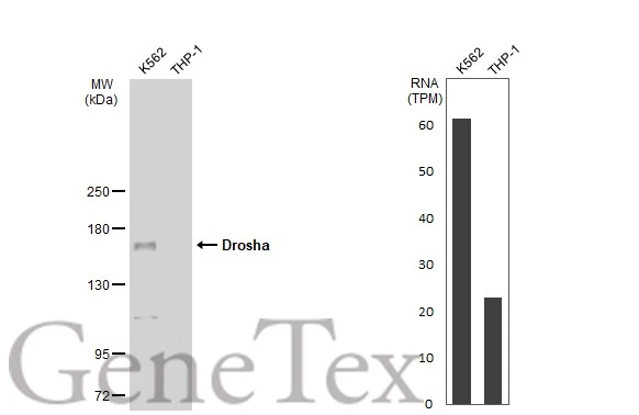
Various whole cell extracts (30 μg) were separated by 5% SDS-PAGE, and the membrane was blotted with Drosha antibody [GT1178] (GTX09511) diluted at 1:1000. The HRP-conjugated anti-rabbit IgG antibody (GTX213110-01) was used to detect the primary antibody. Corresponding RNA expression data for the same cell lines are based on Human Protein Atlas program.
![ICC/IF analysis of HeLa cells using GTX09511 Drosha antibody [GT1178]. Orange: Primary antibody Blue: DAPI](https://www.genetex.com/upload/website/prouct_img/normal/GTX09511/GTX09511_20191101_AP_002_124_w_23053123_809.webp "ICC/IF analysis of HeLa cells using GTX09511 Drosha antibody [GT1178]. Orange: Primary antibody Blue: DAPI")
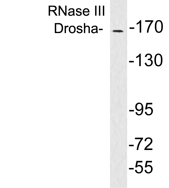
![WB analysis of various samples using GTX09511 Drosha antibody [GT1178]. Dilution : 1:1000 Loading : 25 μg](https://www.genetex.com/upload/website/prouct_img/normal/GTX09511/GTX09511_20200508_WB_w_23053123_229.webp "WB analysis of various samples using GTX09511 Drosha antibody [GT1178]. Dilution : 1:1000 Loading : 25 μg")
Various whole cell extracts (30 μg) were separated by 5% SDS-PAGE, and the membrane was blotted with Drosha antibody [GT1178] (GTX09511) diluted at 1:1000. The HRP-conjugated anti-rabbit IgG antibody (GTX213110-01) was used to detect the primary antibody. Corresponding RNA expression data for the same cell lines are based on Human Protein Atlas program.
Drosha antibody [GT1178]
GTX09511
ApplicationsImmunoFluorescence, Western Blot, ImmunoCytoChemistry
Product group Antibodies
ReactivityHuman
TargetDROSHA
Overview
- SupplierGeneTex
- Product NameDrosha antibody [GT1178]
- Delivery Days Customer9
- Application Supplier NoteWB: 1:500 - 1:2000. ICC/IF: 1:50 - 1:200. *Optimal dilutions/concentrations should be determined by the researcher.Not tested in other applications.
- ApplicationsImmunoFluorescence, Western Blot, ImmunoCytoChemistry
- CertificationResearch Use Only
- ClonalityMonoclonal
- Clone IDGT1178
- ConjugateUnconjugated
- Gene ID29102
- Target nameDROSHA
- Target descriptiondrosha ribonuclease III
- Target synonymsETOHI2, HSA242976, RANSE3L, RN3, RNASE3L, RNASEN, ribonuclease 3, RNase III, drosha, double-stranded RNA-specific endoribonuclease, nuclear RNase III Drosha, p241, putative protein p241 which interacts with transcription factor Sp1, putative ribonuclease III, ribonuclease type III, nuclear
- HostRabbit
- IsotypeIgG
- Protein IDQ9NRR4
- Protein NameRibonuclease 3
- Scientific DescriptionThis gene encodes a ribonuclease (RNase) III double-stranded RNA-specific ribonuclease and subunit of the microprocessor protein complex, which catalyzes the initial processing step of microRNA (miRNA) synthesis. The encoded protein cleaves the stem loop structure from the primary microRNA (pri-miRNA) in the nucleus, yielding the precursor miRNA (pre-miRNA), which is then exported to the cytoplasm for further processing. In a hmuan cell line lacking a functional copy of this gene, canonical miRNA synthesis is reduced. Somatic mutations in this gene have been observed in hmuan patients with kidney cancer. [provided by RefSeq, Sep 2016]
- ReactivityHuman
- Storage Instruction-20°C or -80°C,2°C to 8°C
- UNSPSC41116161
Datasheet
Related products
Product group Antibodies
ApplicationsWestern Blot, ELISA
ReactivityHuman, Mouse
- SizePrice
Product group Antibodies

DROSHA / RNASEN AntibodyLS-C830025
ApplicationsELISA, ImmunoHistoChemistry
ReactivityHuman, Mouse
TargetDROSHA
- SizePrice

Product group Antibodies
Anti-DROSHA Antibody Picoband(r)A00111-3-CARRIER-FREE
ApplicationsFlow Cytometry, Western Blot, ELISA
ReactivityHuman, Mouse
TargetDROSHA
- SizePrice
Product group Antibodies
Drosha Recombinant Antibody, Biotin ConjugatedBSM-61492R-BIOTIN
ApplicationsWestern Blot, ImmunoHistoChemistry, ImmunoHistoChemistry Frozen, ImmunoHistoChemistry Paraffin
ReactivityHuman
TargetDROSHA
- SizePrice
Product group Antibodies
ApplicationsWestern Blot
TargetDROSHA
- SizePrice
Product group Antibodies
DROSHA AntibodyCSB-PA352154
ApplicationsWestern Blot, ELISA, ImmunoHistoChemistry
ReactivityHuman, Mouse
TargetDROSHA
- SizePrice
Product group Antibodies
Drosha antibodyGTX56186
ApplicationsWestern Blot
ReactivityHuman, Mouse, Rat
TargetDROSHA
- SizePrice