

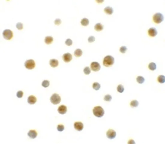
ICC/IF analysis of HeLa cells using GTX31433 DYRK1A antibody. Working concentration : 10 μg/ml
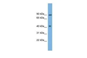
 1 and (B) 2 μg/ml")
 1 and (B) 2 μg/ml")
ICC/IF analysis of HeLa cells using GTX31433 DYRK1A antibody. Working concentration : 10 μg/ml
DYRK1A antibody
GTX31433
ApplicationsImmunoFluorescence, Western Blot, ELISA, ImmunoCytoChemistry
Product group Antibodies
ReactivityHuman, Mouse, Rat
TargetDYRK1A
Overview
- SupplierGeneTex
- Product NameDYRK1A antibody
- Delivery Days Customer9
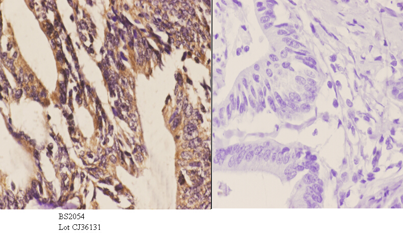
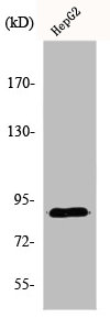
- Application Supplier NoteWB: 1 - 2 microg/mL. ICC/IF: 10 microg/mL. *Optimal dilutions/concentrations should be determined by the researcher.Not tested in other applications.
- ApplicationsImmunoFluorescence, Western Blot, ELISA, ImmunoCytoChemistry
- CertificationResearch Use Only
- ClonalityPolyclonal
- Concentration1 mg/ml
- ConjugateUnconjugated
- Gene ID1859
- Target nameDYRK1A
- Target descriptiondual specificity tyrosine phosphorylation regulated kinase 1A
- Target synonymsDYRK, DYRK1, HP86, MNB, MNBH, MRD7, dual specificity tyrosine-phosphorylation-regulated kinase 1A, MNB/DYRK protein kinase, dual specificity YAK1-related kinase, dual specificity tyrosine-(Y)-phosphorylation regulated kinase 1A, mnb protein kinase homolog hp86, protein kinase minibrain homolog, serine/threonine kinase MNB, serine/threonine-specific protein kinase
- HostRabbit
- IsotypeIgG
- Protein IDQ13627
- Protein NameDual specificity tyrosine-phosphorylation-regulated kinase 1A
- Scientific DescriptionThis gene encodes a member of the Dual-specificity tyrosine phosphorylation-regulated kinase (DYRK) family. This member contains a nuclear targeting signal sequence, a protein kinase domain, a leucine zipper motif, and a highly conservative 13-consecutive-histidine repeat. It catalyzes its autophosphorylation on serine/threonine and tyrosine residues. It may play a significant role in a signaling pathway regulating cell proliferation and may be involved in brain development. This gene is a homolog of Drosophila mnb (minibrain) gene and rat Dyrk gene. It is localized in the Down syndrome critical region of chromosome 21, and is considered to be a strong candidate gene for learning defects associated with Down syndrome. Alternative splicing of this gene generates several transcript variants differing from each other either in the 5 UTR or in the 3 coding region. These variants encode at least five different isoforms. [provided by RefSeq, Jul 2008]
- ReactivityHuman, Mouse, Rat
- Storage Instruction-20°C or -80°C,2°C to 8°C
- UNSPSC41116161
Datasheet
Related products
Product group Antibodies
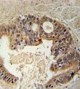
ApplicationsImmunoFluorescence, Western Blot, ImmunoHistoChemistry
ReactivityHuman, Mouse, Rat
- SizePrice

Product group Antibodies
Anti-DYRK1A Antibody Picoband(r)A00878-1-CARRIER-FREE
ApplicationsImmunoFluorescence, Western Blot, ELISA, ImmunoCytoChemistry
ReactivityHuman, Rat
TargetDYRK1A
- SizePrice
Product group Antibodies
Anti-DYRK1A Antibody144-00595
ApplicationsWestern Blot
ReactivityHuman, Mouse, Rat
TargetDYRK1A
- SizePrice
Product group Antibodies
DYRK1A AntibodyCSB-PA002232
ApplicationsImmunoFluorescence, Western Blot, ELISA, ImmunoHistoChemistry
ReactivityHuman, Mouse, Rat
TargetDYRK1A
- SizePrice
Product group Antibodies
References

Goat anti-DYRK1AEB11483
ApplicationsImmunoPrecipitation, Western Blot, ELISA
ReactivityBovine, Canine, Human, Mouse, Porcine, Rat
TargetDYRK1A
- SizePrice
Product group Antibodies
ApplicationsImmunoPrecipitation, Western Blot, ImmunoCytoChemistry, ImmunoHistoChemistry
ReactivityMouse, Porcine, Rat
TargetDYRK1A
- SizePrice
Product group Antibodies

DYRK / DYRK1A AntibodyLS-C408229
ApplicationsWestern Blot
ReactivityHuman, Mouse, Rat
TargetDYRK1A
- SizePrice
Product group Antibodies
DYRK1A antibody, N-termGTX81398
ApplicationsWestern Blot, ImmunoHistoChemistry, ImmunoHistoChemistry Paraffin
ReactivityHuman, Mouse
TargetDYRK1A
- SizePrice
Product group Antibodies
DYRK1A antibody, N-termGTX46524
ApplicationsWestern Blot
ReactivityHuman
TargetDYRK1A
- SizePrice