

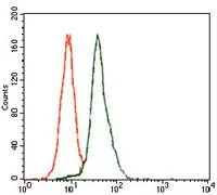
FACS analysis of HeLa cells using GTX60615 E2F1 antibody [8G9]. Green : E2F1 Red : negative control
![IHC-P analysis of rectum cancer tissue using GTX60615 E2F1 antibody [8G9].](https://www.genetex.com/upload/website/prouct_img/normal/GTX60615/GTX60615_20170912_IHC-P_1_w_23061123_723.webp "IHC-P analysis of rectum cancer tissue using GTX60615 E2F1 antibody [8G9].")
![ELISA analysis of antigen using GTX60615 E2F1 antibody [8G9].
Black : Control antigen 100ng
Purple : Antigen 10ng
Blue : Antigen 50ng
Red : Antigen 100ng](https://www.genetex.com/upload/website/prouct_img/normal/GTX60615/GTX60615_20170912_ELISA_w_23061123_554.webp "ELISA analysis of antigen using GTX60615 E2F1 antibody [8G9].
Black : Control antigen 100ng
Purple : Antigen 10ng
Blue : Antigen 50ng
Red : Antigen 100ng")
![WB analysis of HeLa (1), SK-N-SH (2), and NIH3T3 (3) cell lysate using GTX60615 E2F1 antibody [8G9].](https://www.genetex.com/upload/website/prouct_img/normal/GTX60615/GTX60615_20170912_WB_w_23061123_692.webp "WB analysis of HeLa (1), SK-N-SH (2), and NIH3T3 (3) cell lysate using GTX60615 E2F1 antibody [8G9].")
![IHC-P analysis of esophageal cancer tissue using GTX60615 E2F1 antibody [8G9].](https://www.genetex.com/upload/website/prouct_img/normal/GTX60615/GTX60615_20170912_IHC-P_w_23061123_735.webp "IHC-P analysis of esophageal cancer tissue using GTX60615 E2F1 antibody [8G9].")
FACS analysis of HeLa cells using GTX60615 E2F1 antibody [8G9]. Green : E2F1 Red : negative control
E2F1 antibody [8G9]
GTX60615
ApplicationsFlow Cytometry, Western Blot, ELISA, ImmunoHistoChemistry, ImmunoHistoChemistry Paraffin
Product group Antibodies
ReactivityHuman, Mouse
TargetE2F1
Overview
- SupplierGeneTex
- Product NameE2F1 antibody [8G9]
- Delivery Days Customer9
- Application Supplier NoteWB: 1/500 - 1/2000. IHC-P: 1/200 - 1/1000. FACS: 1/200 - 1/400. ELISA: 1/10000. *Optimal dilutions/concentrations should be determined by the researcher.Not tested in other applications.
- ApplicationsFlow Cytometry, Western Blot, ELISA, ImmunoHistoChemistry, ImmunoHistoChemistry Paraffin
- CertificationResearch Use Only
- ClonalityMonoclonal
- Clone ID8G9
- Concentration1 mg/ml
- ConjugateUnconjugated
- Gene ID1869
- Target nameE2F1
- Target descriptionE2F transcription factor 1
- Target synonymsE2F-1, RBAP1, RBBP3, RBP3, transcription factor E2F1, PBR3, PRB-binding protein E2F-1, RBAP-1, RBBP-3, retinoblastoma-associated protein 1, retinoblastoma-binding protein 3, retinoblastoma-binding protein 3 (E2F-like)
- HostMouse
- IsotypeIgG1
- Protein IDQ01094
- Protein NameTranscription factor E2F1
- Scientific DescriptionThe protein encoded by this gene is a member of the E2F family of transcription factors. The E2F family plays a crucial role in the control of cell cycle and action of tumor suppressor proteins and is also a target of the transforming proteins of small DNA tumor viruses. The E2F proteins contain several evolutionally conserved domains found in most members of the family. These domains include a DNA binding domain, a dimerization domain which determines interaction with the differentiation regulated transcription factor proteins (DP), a transactivation domain enriched in acidic amino acids, and a tumor suppressor protein association domain which is embedded within the transactivation domain. This protein and another 2 members, E2F2 and E2F3, have an additional cyclin binding domain. This protein binds preferentially to retinoblastoma protein pRB in a cell-cycle dependent manner. It can mediate both cell proliferation and p53-dependent/independent apoptosis. [provided by RefSeq, Jul 2008]
- ReactivityHuman, Mouse
- Storage Instruction-20°C or -80°C,2°C to 8°C
- UNSPSC12352203
Datasheet
Related products
Product group Antibodies

Transcription factor E2F1 AntibodyABX433397
ApplicationsWestern Blot, ELISA
- SizePrice
Product group Antibodies
Anti-E2F1 Antibody144-64611
ApplicationsImmunoFluorescence, Western Blot
ReactivityHuman, Mouse, Rat
TargetE2F1
- SizePrice

Product group Antibodies
Anti-E2F1 Antibody Picoband(r)A00257-2-CARRIER-FREE
ApplicationsFlow Cytometry, Western Blot, ELISA
ReactivityHuman, Mouse, Rat
TargetE2F1
- SizePrice
![WB analysis of MCF7 untreated (lane 1) or treated with 10μM etoposide for 16 hrs (lane 2 and 3) using GTX00679 E2F1 (phospho Ser364) antibody [#2]. Lane 1 and 2 : IP with pantropic anti-E2F1 antibody followed by WB with GTX00679 Lane 3 : Cell lysate analyzed by WB with GTX00679](https://www.genetex.com/upload/website/prouct_img/normal/GTX00679/GTX00679_20191104_WB_w_23053121_640.webp)
Product group Antibodies
ApplicationsWestern Blot, ELISA
ReactivityHuman
TargetE2F1
- SizePrice
![Various whole cell extracts (30 μg) were separated by 10% SDS-PAGE, and the membrane was blotted with E2F1 antibody [GT1167] (GTX09015) diluted at 1:500. The HRP-conjugated anti-rabbit IgG antibody (GTX213110-01) was used to detect the primary antibody.](https://www.genetex.com/upload/website/prouct_img/normal/GTX09015/GTX09015_40000000058_20200306_WB_w_23053123_653.webp)
Product group Antibodies
E2F1 antibody [GT1167]GTX09015
ApplicationsWestern Blot
ReactivityHuman, Mouse, Rat
TargetE2F1
- SizePrice
![Non-transfected (–) and transfected (+) 293T whole cell extracts (30 μg) were separated by 10% SDS-PAGE, and the membrane was blotted with E2F1 antibody [N1N3] (GTX101235) diluted at 1:500. The HRP-conjugated anti-rabbit IgG antibody (GTX213110-01) was used to detect the primary antibody.](https://www.genetex.com/upload/website/prouct_img/normal/GTX101235/GTX101235_39812_20181221_WB_B_w_23060100_187.webp)
Product group Antibodies
References
E2F1 antibody [N1N3]GTX101235
ApplicationsImmunoFluorescence, ImmunoPrecipitation, Western Blot, ImmunoCytoChemistry
ReactivityHuman
TargetE2F1
- SizePrice
Product group Antibodies
Anti-E2F1Y058486
ApplicationsWestern Blot, ImmunoHistoChemistry
ReactivityHuman
- SizePrice
![IP analysis of HeLa whole cell lysate using GTX66839 E2F1 antibody [4G8].](https://www.genetex.com/upload/website/prouct_img/normal/GTX66839/GTX66839_20220818_IP_22081801_290.webp)
Product group Antibodies
E2F1 antibody [4G8]GTX66839
ApplicationsImmunoFluorescence, ImmunoPrecipitation, Western Blot, ImmunoCytoChemistry
ReactivityHuman, Mouse, Rat
TargetE2F1
- SizePrice
![Mouse tissue extract (50 μg) was separated by 10% SDS-PAGE, and the membrane was blotted with E2F1 antibody [17E2] (GTX70154) diluted at 1:1000. The HRP-conjugated anti-mouse IgG antibody (GTX213111-01) was used to detect the primary antibody.](https://www.genetex.com/upload/website/prouct_img/normal/GTX70154/GTX70154_44482_20211112_WB_M_spleen_w_23061221_533.webp)
Product group Antibodies
E2F1 antibody [17E2]GTX70154
ApplicationsImmunoPrecipitation, Western Blot, ImmunoHistoChemistry, ImmunoHistoChemistry Paraffin
ReactivityHuman, Mouse
TargetE2F1
- SizePrice
Product group Antibodies
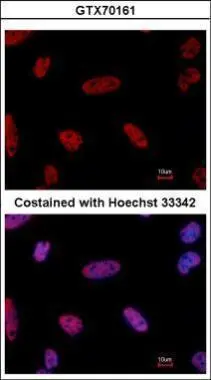
E2F1 antibody [10D1 2B5]GTX70161
ApplicationsImmunoFluorescence, ImmunoPrecipitation, Western Blot, ImmunoCytoChemistry
ReactivityHuman
TargetE2F1
- SizePrice