

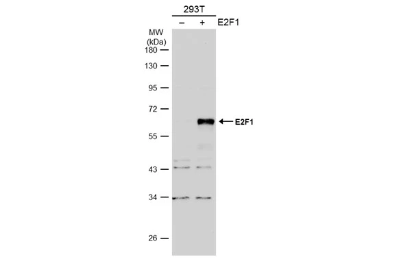
Non-transfected (–) and transfected (+) 293T whole cell extracts (30 μg) were separated by 10% SDS-PAGE, and the membrane was blotted with E2F1 antibody [N1N3] (GTX101235) diluted at 1:500. The HRP-conjugated anti-rabbit IgG antibody (GTX213110-01) was used to detect the primary antibody.
![E2F1 antibody [N1N3] detects E2F1 protein at nucleus by immunofluorescent analysis. Sample: HeLa cells were fixed in 4% paraformaldehyde at RT for 15 min. Green: E2F1 protein stained by E2F1 antibody [N1N3] (GTX101235) diluted at 1:500. Red: Phalloidin, a cytoskeleton marker, diluted at 1:200. Scale bar = 10 μm.](https://www.genetex.com/upload/website/prouct_img/normal/GTX101235/GTX101235_39812_20151216_IFA_w_23060100_370.webp "E2F1 antibody [N1N3] detects E2F1 protein at nucleus by immunofluorescent analysis. Sample: HeLa cells were fixed in 4% paraformaldehyde at RT for 15 min. Green: E2F1 protein stained by E2F1 antibody [N1N3] (GTX101235) diluted at 1:500. Red: Phalloidin, a cytoskeleton marker, diluted at 1:200. Scale bar = 10 μm.")
Non-transfected (–) and transfected (+) 293T whole cell extracts (30 μg) were separated by 10% SDS-PAGE, and the membrane was blotted with E2F1 antibody [N1N3] (GTX101235) diluted at 1:500. The HRP-conjugated anti-rabbit IgG antibody (GTX213110-01) was used to detect the primary antibody.
E2F1 antibody [N1N3]
GTX101235
ApplicationsImmunoFluorescence, ImmunoPrecipitation, Western Blot, ImmunoCytoChemistry
Product group Antibodies
ReactivityHuman
TargetE2F1
Overview
- SupplierGeneTex
- Product NameE2F1 antibody [N1N3]
- Delivery Days Customer9
- Application Supplier NoteWB: 1:500-1:3000. ICC/IF: 1:100-1:1000. IP: 1:100-1:500. *Optimal dilutions/concentrations should be determined by the researcher.Not tested in other applications.
- ApplicationsImmunoFluorescence, ImmunoPrecipitation, Western Blot, ImmunoCytoChemistry
- CertificationResearch Use Only
- ClonalityPolyclonal
- Concentration1 mg/ml
- ConjugateUnconjugated
- Gene ID1869
- Target nameE2F1
- Target descriptionE2F transcription factor 1
- Target synonymsE2F-1, RBAP1, RBBP3, RBP3, transcription factor E2F1, PBR3, PRB-binding protein E2F-1, RBAP-1, RBBP-3, retinoblastoma-associated protein 1, retinoblastoma-binding protein 3, retinoblastoma-binding protein 3 (E2F-like)
- HostRabbit
- IsotypeIgG
- Protein IDQ01094
- Protein NameTranscription factor E2F1
- Scientific DescriptionThe protein encoded by this gene is a member of the E2F family of transcription factors. The E2F family plays a crucial role in the control of cell cycle and action of tumor suppressor proteins and is also a target of the transforming proteins of small DNA tumor viruses. The E2F proteins contain several evolutionally conserved domains found in most members of the family. These domains include a DNA binding domain, a dimerization domain which determines interaction with the differentiation regulated transcription factor proteins (DP), a transactivation domain enriched in acidic amino acids, and a tumor suppressor protein association domain which is embedded within the transactivation domain. This protein and another 2 members, E2F2 and E2F3, have an additional cyclin binding domain. This protein binds preferentially to retinoblastoma protein pRB in a cell-cycle dependent manner. It can mediate both cell proliferation and p53-dependent/independent apoptosis. [provided by RefSeq]
- ReactivityHuman
- Storage Instruction-20°C or -80°C,2°C to 8°C
- UNSPSC41116161
Datasheet
Related products
Product group Antibodies
Anti-E2F1 AntibodyA83673
ApplicationsImmunoFluorescence, ELISA
ReactivityDrosophila
- SizePrice
Product group Antibodies
Anti-E2F1 Antibody144-64611
ApplicationsImmunoFluorescence, Western Blot
ReactivityHuman, Mouse, Rat
TargetE2F1
- SizePrice
Product group Antibodies

Transcription factor E2F1 AntibodyABX433397
ApplicationsWestern Blot, ELISA
- SizePrice
Product group Antibodies

E2F1 AntibodyLS-C812948
ApplicationsWestern Blot, ELISA
ReactivityHuman, Rat
TargetE2F1
- SizePrice

Product group Antibodies
Anti-E2F1 Antibody Picoband(r)A00257-2-CARRIER-FREE
ApplicationsFlow Cytometry, Western Blot, ELISA
ReactivityHuman, Mouse, Rat
TargetE2F1
- SizePrice
Product group Antibodies
References
E2F1 Polyclonal AntibodyBS-0599R
ApplicationsFlow Cytometry, ImmunoFluorescence, Western Blot, ELISA, ImmunoCytoChemistry, ImmunoHistoChemistry, ImmunoHistoChemistry Frozen, ImmunoHistoChemistry Paraffin
ReactivityBovine, Chicken, Equine, Human, Mouse, Porcine, Rabbit, Rat, Sheep
TargetE2F1
- SizePrice
Product group Antibodies
E2F1 AntibodyCSB-PA002235
ApplicationsWestern Blot, ELISA, ImmunoHistoChemistry
ReactivityHuman, Mouse
TargetE2F1
- SizePrice
Product group Antibodies

ApplicationsWestern Blot, ELISA
ReactivityHuman
TargetE2F1
- SizePrice
Product group Antibodies
E2F1 Polyclonal AntibodyCAC14530
ApplicationsImmunoPrecipitation, Western Blot, ELISA, ImmunoHistoChemistry
ReactivityMouse
TargetE2F1
- SizePrice
Product group Antibodies
E2F1 antibodyGTX14770
ApplicationsWestern Blot, ELISA
ReactivityHuman, Mouse, Rat
TargetE2F1
- SizePrice