

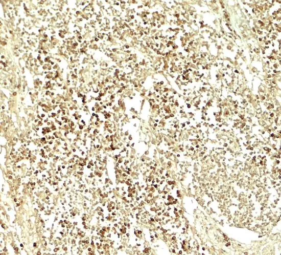
IHC-P analysis of human lymph node tissue using GTX31589 E2F3 antibody. Working concentration : 5 μg/ml


IHC-P analysis of human lymph node tissue using GTX31589 E2F3 antibody. Working concentration : 5 μg/ml
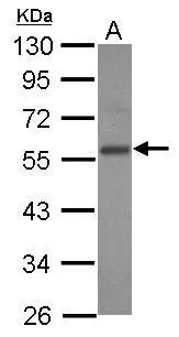
E2F3 antibody
GTX31589
ApplicationsWestern Blot, ELISA, ImmunoHistoChemistry, ImmunoHistoChemistry Paraffin
Product group Antibodies
ReactivityHuman, Mouse
TargetE2F3
Overview
- SupplierGeneTex
- Product NameE2F3 antibody
- Delivery Days Customer9
- Application Supplier NoteWB: 1 - 2 microg/mL. IHC-P: 5 microg/mL. *Optimal dilutions/concentrations should be determined by the researcher.Not tested in other applications.
- ApplicationsWestern Blot, ELISA, ImmunoHistoChemistry, ImmunoHistoChemistry Paraffin
- CertificationResearch Use Only
- ClonalityPolyclonal
- Concentration1 mg/ml
- ConjugateUnconjugated
- Gene ID1871
- Target nameE2F3
- Target descriptionE2F transcription factor 3
- Target synonymsE2F-3, transcription factor E2F3
- HostRabbit
- IsotypeIgG
- Protein IDO00716
- Protein NameTranscription factor E2F3
- Scientific DescriptionThis gene encodes a member of a small family of transcription factors that function through binding of DP interaction partner proteins. The encoded protein recognizes a specific sequence motif in DNA and interacts directly with the retinoblastoma protein (pRB) to regulate the expression of genes involved in the cell cycle. Altered copy number and activity of this gene have been observed in a number of human cancers. There are pseudogenes for this gene on chromosomes 2 and 17. Alternative splicing results in multiple transcript variants. [provided by RefSeq, Mar 2013]
- ReactivityHuman, Mouse
- Storage Instruction-20°C or -80°C,2°C to 8°C
- UNSPSC41116161
Datasheet
Related products
Product group Antibodies
E2F3 AntibodyCSB-PA007342LA01HU
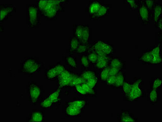
ApplicationsImmunoFluorescence, ELISA
ReactivityHuman
- SizePrice
Product group Antibodies
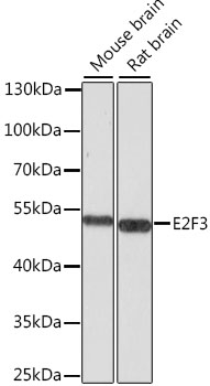
Anti-E2F3 AntibodyA12305
ApplicationsWestern Blot
ReactivityMouse, Rat
- SizePrice

Product group Antibodies
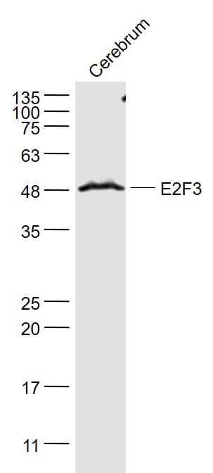
Anti-E2F3 Antibody Picoband(r)A03068-2-CARRIER-FREE
ApplicationsWestern Blot
ReactivityHuman, Mouse, Rat
TargetE2F3
- SizePrice
Product group Antibodies

Goat anti-E2F3 (mouse)EB09935
ApplicationsWestern Blot, ELISA
ReactivityHuman, Mouse
TargetE2F3
- SizePrice

Product group Antibodies
Anti-E2F3 AntibodyHPA029779
ApplicationsImmunoCytoChemistry
ReactivityHuman
TargetE2F3
- SizePrice
Product group Antibodies

E2F3 AntibodyLS-C410345
ApplicationsWestern Blot, ImmunoHistoChemistry
ReactivityHuman, Mouse
TargetE2F3
- SizePrice
Product group Antibodies
References
E2F3 Polyclonal AntibodyBS-1722R
ApplicationsImmunoFluorescence, Western Blot, ELISA, ImmunoCytoChemistry, ImmunoHistoChemistry, ImmunoHistoChemistry Frozen, ImmunoHistoChemistry Paraffin
ReactivityBovine, Canine, Equine, Human, Mouse, Porcine, Rabbit, Rat
TargetE2F3
- SizePrice
Product group Antibodies
E2F3 antibody [N2C3]GTX102302
ApplicationsWestern Blot
ReactivityHuman
TargetE2F3
- SizePrice
![WB analysis of 293T cell lysates using GTX11843 E2F3 antibody [PG30].
Dilution : Lane1 : 4 μg/mL Lane2 : 8 μg/mL](https://www.genetex.com/upload/website/prouct_img/normal/GTX11843/GTX11843_20221006_WB_22100518_247.webp)
Product group Antibodies
References
E2F3 antibody [PG30]GTX11843
ApplicationsImmunoPrecipitation, Western Blot
ReactivityHuman
TargetE2F3
- SizePrice
Product group Antibodies
Anti-E2F3 Antibody144-66688
ApplicationsWestern Blot
ReactivityHuman, Mouse, Rat
TargetE2F3
- SizePrice