Bio-Connect
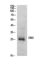
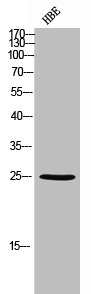
Gel: 12%SDS-PAGE Lysate: 40 microg Lane 1-2: Mouse kidney tissue 293T cell lysates Primary antibody: TA365338 (EBI3 Antibody) at dilution 1/400 Secondary antibody: Goat anti rabbit IgG at 1/5000 dilution Exposure time: 3 minutes

EBI3 Rabbit Polyclonal Antibody

TA365338
OriGene
ApplicationsWestern Blot, ImmunoHistoChemistry
Product group Antibodies
TargetEBI3
100 ul
Sign in to order and to see your custom pricing.
Large volume orders?
Order with a bulk request

Overview

  • Supplier
    OriGene
  • Product Name
    EBI3 Rabbit Polyclonal Antibody
  • Delivery Days Customer
    14
  • Applications
    Western Blot, ImmunoHistoChemistry
  • Certification
    Research Use Only
  • Clonality
    Polyclonal
  • Gene ID10148
  • Target name
    EBI3
  • Target description
    Epstein-Barr virus induced 3
  • Target synonyms
    cytokine receptor; EBV-induced gene 3 protein; Epstein-Barr virus induced gene 3; epstein-Barr virus-induced gene 3 protein; IL27 subunit; IL-27 subunit beta; IL27B; IL-27B; IL35 subunit; IL35B; interleukin-27 subunit beta
  • Host
    Rabbit
  • Isotype
    IgG
  • Protein IDQ14213
  • Protein Name
    Interleukin-27 subunit beta
  • Scientific Description
    EBI3 rabbit polyclonal antibody
  • Storage Instruction
    -20°C
  • UNSPSC
    12352203

Related products

Product group Antibodies
Bioss Antibodies
ApplicationsWestern Blot
TargetEBI3
  • SizePrice
Figure 1. Western blot analysis of EBI3 using anti-EBI3 antibody (M07190-2). Electrophoresis was performed on a 5-20% SDS-PAGE gel at 70V (Stacking gel) / 90V (Resolving gel) for 2-3 hours. The sample well of each lane was loaded with 30 ug of sample under reducing conditions. Lane 1: human K562 whole cell lysates, Lane 2: human U20S whole cell lysates, Lane 3: human Caco-2 whole cell lysates, Lane 4: rat spleen tissue lysates, Lane 5: rat thymus tissue lysates, Lane 7: mouse spleen tissue lysates, Lane 8: mouse thymus tissue lysates. After electrophoresis, proteins were transferred to a nitrocellulose membrane at 150 mA for 50-90 minutes. Blocked the membrane with 5% non-fat milk/TBS for 1.5 hour at RT. The membrane was incubated with rabbit anti-EBI3 antigen affinity purified monoclonal antibody (Catalog # M07190-2) at 1:500 overnight at 4°C, then washed with TBS-0.1%Tween 3 times with 5 minutes each and probed with a goat anti-rabbit IgG-HRP secondary antibody at a dilution of 1:500 for 1.5 hour at RT. The signal is developed using an Enhanced Chemiluminescent detection (ECL) kit (Catalog # EK1002) with Tanon 5200 system. A specific band was detected for EBI3 at approximately 30 kDa. The expected band size for EBI3 is at 25,30 kDa.
Product group Antibodies
Boster Bio
ApplicationsWestern Blot
TargetEBI3
  • SizePrice
Product group Antibodies
RayBiotech
ApplicationsWestern Blot
TargetEBI3
  • SizePrice
Product group Antibodies
Biosynth
  • SizePrice
Anti-EBI3 Antibody
Product group Antibodies
Antibodies.com
ApplicationsWestern Blot, ELISA
  • SizePrice
Anti-human EBI3 mAb (MT23A14), unconjugated
Product group Antibodies
Mabtech
ApplicationsELISA, ELISpot Assay
TargetEBI3
  • SizePrice
Product group Antibodies
MedChem Express
TargetEBI3
  • SizePrice
Western Blot analysis of HBE cells using Ebi3 Polyclonal Antibody
Product group Antibodies
Cusabio
EBI3 AntibodyCSB-PA006315
ApplicationsWestern Blot, ELISA
ReactivityHuman, Mouse
TargetEBI3
  • SizePrice
Product group Antibodies
LifeSpan BioSciences
ApplicationsImmunoHistoChemistry
TargetEBI3
  • SizePrice
Product group Antibodies
Biomatik
ApplicationsWestern Blot, ELISA, ImmunoHistoChemistry
TargetEBI3
  • SizePrice